. 2013 Feb 1;288(5):3305-11.
doi: 10.1074/jbc.M112.387712. Epub 2012 Dec 12.

Watching the bacteriophage N4 RNA polymerase transcription by time-dependent soak-trigger-freeze X-ray crystallography

Affiliations

Watching the bacteriophage N4 RNA polymerase transcription by time-dependent soak-trigger-freeze X-ray crystallography

Ritwika S Basu et al. J Biol Chem. .

Abstract

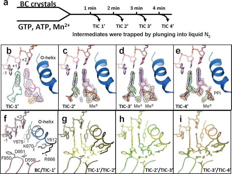
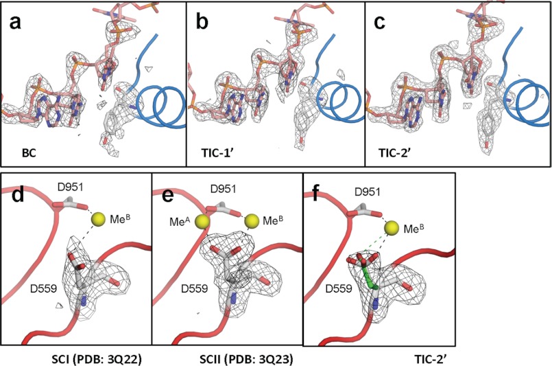
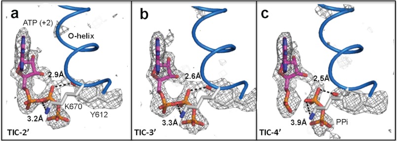
The challenge for structural biology is to understand atomic-level macromolecular motions during enzymatic reaction. X-ray crystallography can reveal high resolution structures; however, one perceived limitation is that it reveals only static views. Here we use time-dependent soak-trigger-freeze X-ray crystallography, namely, soaking nucleotide and divalent metal into the bacteriophage RNA polymerase (RNAP)-promoter DNA complex crystals to trigger the nucleotidyl transfer reaction and freezing crystals at different time points, to capture real-time intermediates in the pathway of transcription. In each crystal structure, different intensities and shapes of electron density maps corresponding to the nucleotide and metal were revealed at the RNAP active site which allow watching the nucleotide and metal bindings and the phosphodiester bond formation in a time perspective. Our study provides the temporal order of substrate assembly and metal co-factor binding at the active site of enzyme which completes our understanding of the two-metal-ion mechanism and fidelity mechanism in single-subunit RNAPs. The nucleotide-binding metal (Me(B)) is coordinated at the active site prior to the catalytic metal (Me(A)). Me(A) coordination is only temporal, established just before and dissociated immediately after phosphodiester bond formation. We captured these elusive intermediates exploiting the slow enzymatic reaction in crystallo. These results demonstrate that the simple time-dependent soak-trigger-freeze X-ray crystallography offers a direct means for monitoring enzymatic reactions.


Figures

FIGURE 1.
FIGURE 1.
Structure of the transcript initiation complex. a, schematic of the transcript initiation. The RNAP and promoter DNA complex is depicted as E. The appearance of intermediates, from TIC-1′ to TIC-4′, isolated in this study as well as SCI, SCII, and PC identified in the previous study (17) are indicated. b, overall structure of the TIC-3′. Each domain and the O-helix of RNAP are denoted by a unique color and labeled. c, sequence of the P2_7c DNA used for crystallization (red, +1 and +2 nucleotide binding sites; gray boxes, disordered in the crystal structures). The position of promoter hairpin is indicated. d, TIC-3′ active site. The amino acid residues involved in nucleotides and metal binding are shown. Nucleotides of +1 (green) and +2 (magenta) are shown. Mn2+ and water are depicted by yellow and cyan spheres, respectively.
FIGURE 2.
FIGURE 2.
Structures of RNAP active site, DNA, nucleotides, and metals during the phosphodiester bond formation. a, scheme of TIC preparations and structure determinations by the time-dependent soak-trigger-freeze X-ray crystallography. b–e, electron density (FoFc, σ cutoff = 4) maps showing nucleotides, 2-mer RNA, pyrophosphate, and metals found in the TIC-1′–TIC-4′ structures. These maps were calculated using the native amplitudes and the phase derived from the final structures without nucleotides, metals, 2-mer RNA, and/or pyrophosphate. In b, the positions of GTP(+1), ATP(+2), and MeB binding sites, which are based on the TIC-3′ structure (d), are indicated by green, magenta, and yellow dashed lines, respectively. f–i, conformational changes of RNAP and DNA during the transcript initiation showing superposition of the BC versus TIC-1′ (f), TIC-1′ versus TIC-2′ (g), TIC-2′ versus TIC-3′ (h), and TIC-3′ versus TIC-4′ (i). BC is colored as in Fig. 1d, and TIC-1′, TIC-2′, TIC-3′, and TIC-4′ are colored in black, yellow, green, and orange, respectively.
FIGURE 3.
FIGURE 3.
Structural transitions of the active site. a–c, FoFc maps (black net, σ cutoff = 3) showing Y-678 and DNA template from −1 to +2 positions superimposed on the final models (sticks and tubes) of BC (a), TIC-1′ (b), and TIC-2′ (c). d–f, FoFc maps (black net, σ cutoff = 3) showing D-559 superimposed on the final models (sticks, tubes, and spheres) of SCI (d), SCII (e), and TIC-2′ (f). Two conformers of D-559 found in the TIC-2′ are shown (green, MeB coordinating form; white, both MeA and MeB coordinating form).
FIGURE 4.
FIGURE 4.
Electron density of Y-612 suggesting proton transfer mechanism. a–c, FoFc maps (black net, σ cutoff = 3) of Y-612 and ATP (+2) superimposed on final models (sticks, Y-612, K-670, and ATP (+2); scheme, O-helix) of TIC-2′ (a), TIC-3′ (b), and TIC-4′ (c). Distances between atoms (black dotted lines) are labeled.

References

    1. Henzler-Wildman K., Kern D. (2007) Dynamic personalities of proteins. Nature 450, 964–972 - PubMed
    1. Bourgeois D., Royant A. (2005) Advances in kinetic protein crystallography. Curr. Opin. Struct. Biol. 15, 538–547 - PubMed
    1. Schmidt M. (2008) in Ultrashort Laser Pulses in Biology and Medicine (Braun M., Gilch P., Zinth W., eds) pp. 201–241, Springer, Berlin
    1. Beese L. S., Steitz T. A. (1991) Structural basis for the 3′-5′ exonuclease activity of Escherichia coli DNA polymerase I: a two metal ion mechanism. EMBO J. 10, 25–33 - PMC - PubMed
    1. Steitz T. A., Steitz J. A. (1993) A general two-metal-ion mechanism for catalytic RNA. Proc. Natl. Acad. Sci. U.S.A. 90, 6498–6502 - PMC - PubMed

Publication types