. 2013 Jan 25;288(4):2893-904.
doi: 10.1074/jbc.M112.411769. Epub 2012 Dec 12.

RapA2 is a calcium-binding lectin composed of two highly conserved cadherin-like domains that specifically recognize Rhizobium leguminosarum acidic exopolysaccharides

Affiliations

RapA2 is a calcium-binding lectin composed of two highly conserved cadherin-like domains that specifically recognize Rhizobium leguminosarum acidic exopolysaccharides

Patricia L Abdian et al. J Biol Chem. .

Abstract

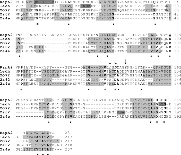
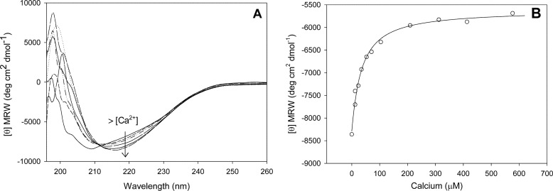
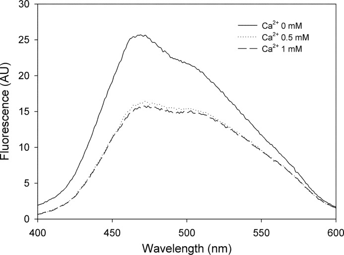
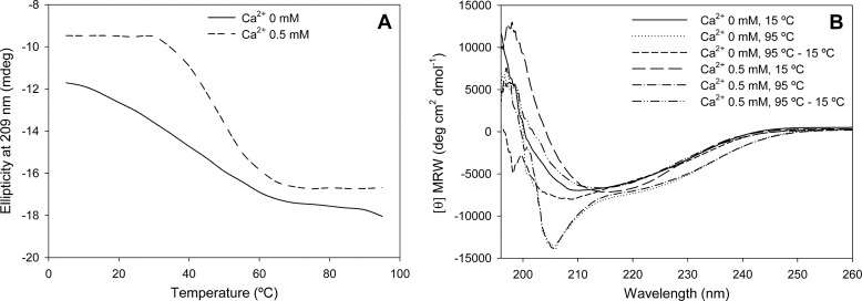
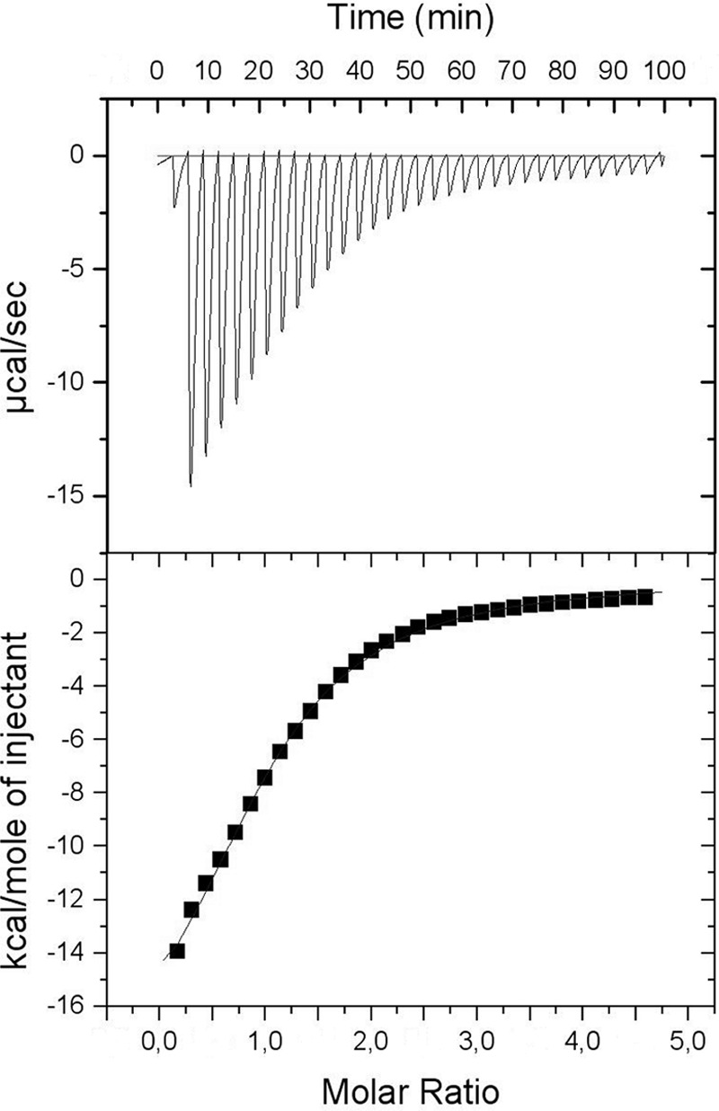
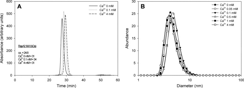
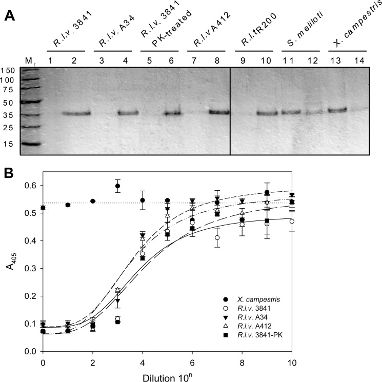
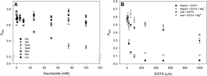
In silico analyses have revealed a conserved protein domain (CHDL) widely present in bacteria that has significant structural similarity to eukaryotic cadherins. A CHDL domain was shown to be present in RapA, a protein that is involved in autoaggregation of Rhizobium cells, biofilm formation, and adhesion to plant roots as shown by us and others. Structural similarity to cadherins suggested calcium-dependent oligomerization of CHDL domains as a mechanistic basis for RapA action. Here we show by circular dichroism spectroscopy, light scattering, isothermal titration calorimetry, and other methods that RapA2 from Rhizobium leguminosarum indeed exhibits a cadherin-like β-sheet conformation and that its proper folding and stability are dependent on the binding of one calcium ion per protein molecule. By further in silico analysis we also reveal that RapA2 consists of two CHDL domains and expand the range of CHDL-containing proteins in bacteria and archaea. However, light scattering assays at various concentrations of added calcium revealed that RapA2 formed neither homo-oligomers nor hetero-oligomers with RapB (a distinct CHDL protein), indicating that RapA2 does not mediate cellular interactions through a cadherin-like mechanism. Instead, we demonstrate that RapA2 interacts specifically with the acidic exopolysaccharides (EPSs) produced by R. leguminosarum in a calcium-dependent manner, sustaining a role of these proteins in the development of the biofilm matrix made of EPS. Because EPS binding by RapA2 can only be attributed to its two CHDL domains, we propose that RapA2 is a calcium-dependent lectin and that CHDL domains in various bacterial and archaeal proteins confer carbohydrate binding activity to these proteins.


Figures

FIGURE 1.
FIGURE 1.
Structural alignment of RapA2 and eukaryotic cadherins. The amino acid sequence of RapA2 was aligned to template sequences according to the MetaServer predictions. The template structures and their PDB entries were: ectodomains 1 and 2 (EC1–2) of mouse E-cadherin (PDB code 1edh), human E-cadherin EC1–2 (PDB 2o72), mouse cadherin-8 EC1–3 (PDB code 2a62), and mouse cadherin-11 EC1–2 (PDB code 2a4e). The predicted RapA2 secondary structure is from SWISS-MODEL, and the template structures were from the Protein Data Bank. Secondary structure elements are shown as a gray background (light gray for β-strands, dark gray for α-helices). Conserved residues are in bold, asterisks denote identical residues, and circles indicate similar residues. The conserved calcium binding motifs in cadherins (DXNDN, DXD and LDRE) are underlined in the sequence of PDB code 1edh. The acidic amino acids of RapA2 mutated to alanine are shown in italics and indicated with arrows.
FIGURE 2.
FIGURE 2.
Effect of calcium on RapA2 secondary structure. A, shown are far-UV CD spectra at increasing calcium concentrations, ranging from 0 to 600 μm. B, shown is molar ellipticity at 208 nm measured at increasing calcium concentrations. MRW, mean residue weight.
FIGURE 3.
FIGURE 3.
ANS binding to RapA2 at increasing calcium concentrations. Intensity of fluorescence emission of ANS when bound to RapA2 at 0, 0.5 or 1 mm calcium is shown. AU, arbitrary units.
FIGURE 4.
FIGURE 4.
Analysis of the effect of calcium on RapA2 thermal stability. A, far-UV CD spectra of RapA2 were recorded at temperatures ranging from 0 to 95 °C. The ellipticity at 209 nm of RapA2 in the absence (apo-form, line) or in the presence of 0.5 mm CaCl2 (holo-form, dashed line) is depicted at different temperatures. B, the reversibility of thermal-induced denaturation of the apo- and holo-forms of RapA2 was determined. Far-UV CD spectra were recorded at 15 °C and 95 °C, and after cooling to 15 °C the samples submitted to 95 °C.
FIGURE 5.
FIGURE 5.
Isotherm of calcium binding to RapA2 measured by ITC. Protein and ligand were prepared in 20 mm Tris-HCl, 150 mm NaCl, pH 8.0. RapA2 (400 μm) in the sample cell was titrated with CaCl2 (10 mm). The upper panel shows the observed heats for each injection of CaCl2 after base-line correction. The lower panel depicts the binding enthalpies versus the calcium/protein molar ratio. The data (■) best fitted to the one set of site binding model. Best-fit parameters are listed in Table 1.
FIGURE 6.
FIGURE 6.
Hydrodynamic properties and diameter distribution of RapA2 at different CaCl2 concentrations. A, shown is molecular mass of RapA2 at various CaCl2 concentrations obtained by size exclusion chromatography and static light scattering. The theoretical and experimentally determined Mr are indicated. B, diameter distribution of RapA2 was measured by DLS at CaCl2 concentrations ranging from 0 to 4 mm.
FIGURE 7.
FIGURE 7.
Interaction of RapA2 with bacterial soluble EPS. A, binding of RapA2 to different soluble polysaccharides was examined. The protein was added to the EPS preparation, and after sedimentation of the EPS by centrifugation at 100,000 × g, the pellet (even numbers) and supernatant fractions (odd numbers) were assayed for the presence of proteins by SDS-PAGE. B, shown is inhibition of the binding of RapA2 to EPS of R. leguminosarum bv. viciae strain 3841 by different soluble EPS preparations. Inhibitory EPS was serially diluted by a factor of 10, starting with a concentration of 100 μg ml−1. R.l.v., R. leguminosarum bv. viciae; R.l.t., R. leguminosarum bv. trifolii; PK, pretreatment with proteinase K.
FIGURE 8.
FIGURE 8.
Inhibitory properties of monosaccharides and EGTA on RapA2-EPS interaction. A, inhibition curves of RapA2 binding to immobilized EPS were obtained by preincubation of the protein with increasing concentrations of the indicated sugars ranging from 0 to 100 mm. B, the effect of EGTA on RapA2 binding to the EPS was determined by preincubation of RapA2 with various concentrations of the chelating agent (0–1 mm). As a control, EGTA was also added to the EPS-coated wells before RapA2 addition. In some cases MgCl2 was added at 500 μm to evaluate its effect on the immobilized EPS in the wells.

References

    1. Downie J. A. (2010) The roles of extracellular proteins, polysaccharides, and signals in the interactions of rhizobia with legume roots. FEMS Microbiol. Rev. 34, 150–170 - PubMed
    1. Rinaudi L. V., Giordano W. (2010) An integrated view of biofilm formation in rhizobia. FEMS Microbiol. Lett. 304, 1–11 - PubMed
    1. Russo D. M., Williams A., Edwards A., Posadas D. M., Finnie C., Dankert M., Downie J. A., Zorreguieta A. (2006) Proteins exported via the PrsD-PrsE type I secretion system and the acidic exopolysaccharide are involved in biofilm formation by Rhizobium leguminosarum. J. Bacteriol. 188, 4474–4486 - PMC - PubMed
    1. Williams A., Wilkinson A., Krehenbrink M., Russo D. M., Zorreguieta A., Downie J. A. (2008) Glucomannan-mediated attachment of Rhizobium leguminosarum to pea root hairs is required for competitive nodule infection. J. Bacteriol. 190, 4706–4715 - PMC - PubMed
    1. Krehenbrink M., Downie J. A. (2008) Identification of protein secretion systems and novel secreted proteins in Rhizobium leguminosarum bv. viciae. BMC Genomics 9, 55. - PMC - PubMed

Publication types

MeSH terms