. 2013 Feb 1;288(5):3469-75.
doi: 10.1074/jbc.M112.413708. Epub 2012 Dec 12.

Purification and characterization of Escherichia coli MreB protein

Affiliations

Purification and characterization of Escherichia coli MreB protein

Pearl Nurse et al. J Biol Chem. .

Abstract

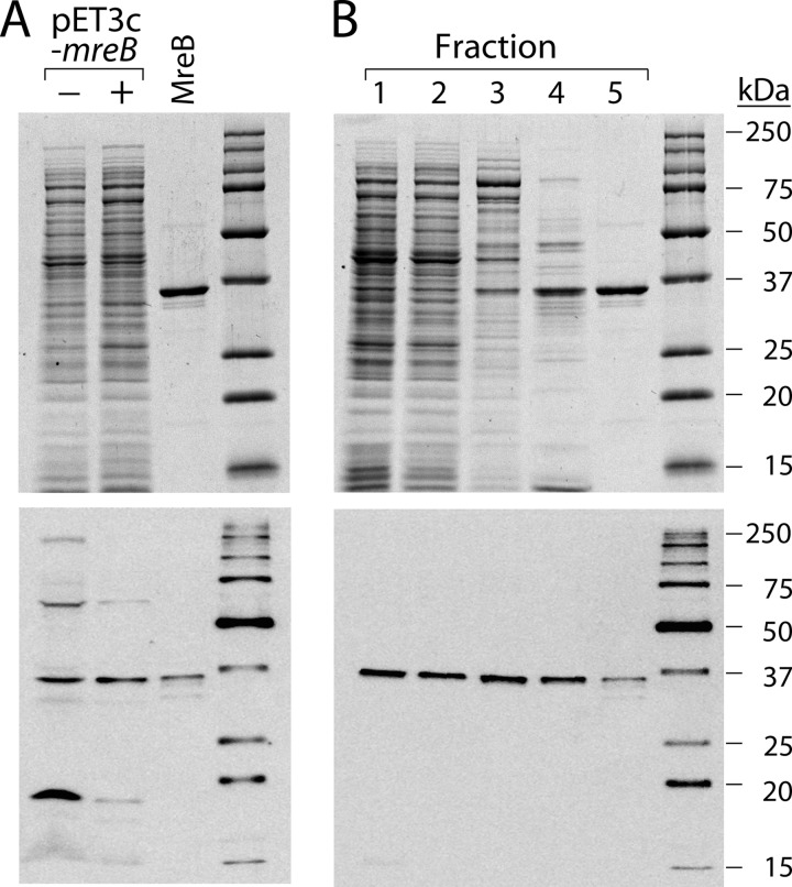
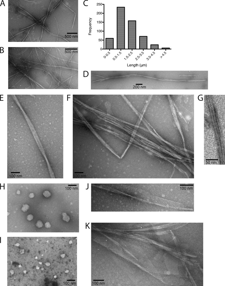
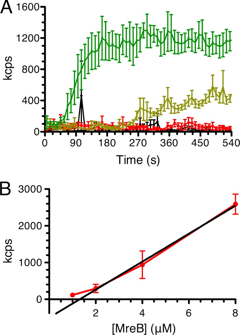
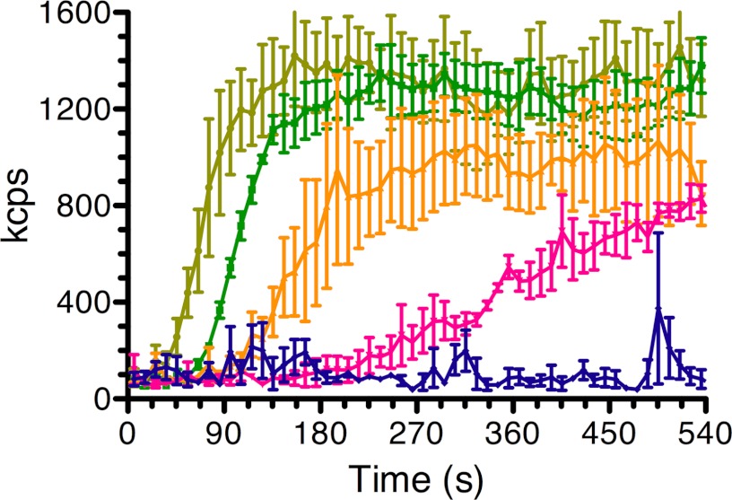
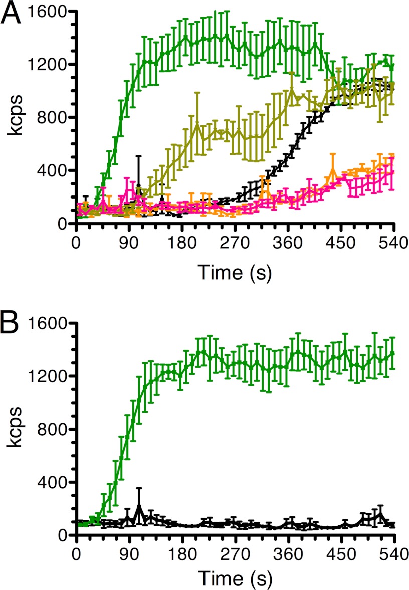
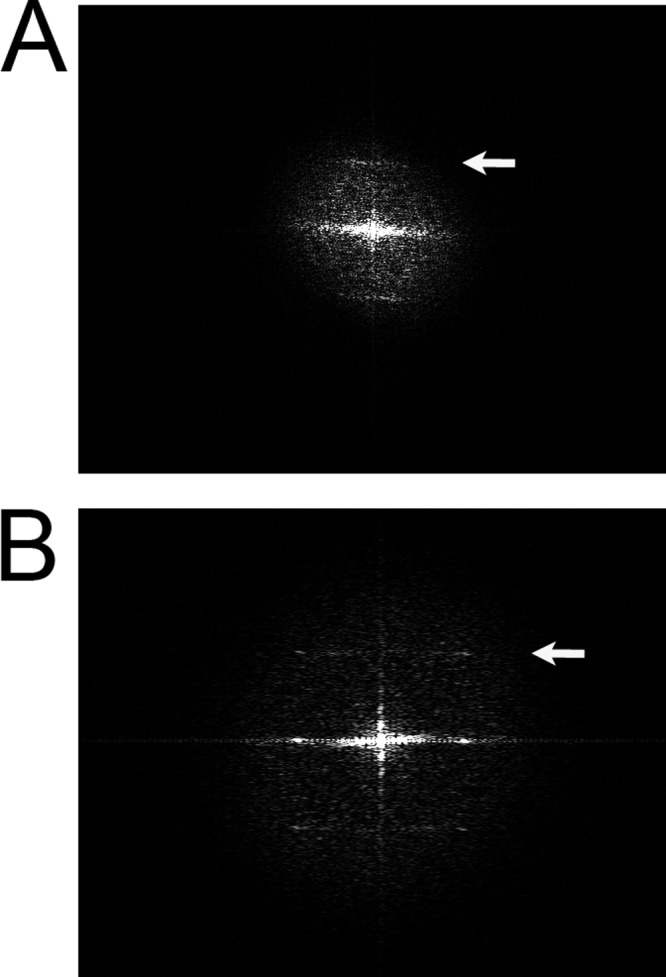
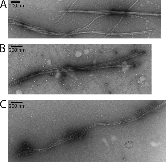
The actin homolog MreB is required in rod-shaped bacteria for maintenance of cell shape and is intimately connected to the holoenzyme that synthesizes the peptidoglycan layer. The protein has been reported variously to exist in helical loops under the cell surface, to rotate, and to move in patches in both directions around the cell surface. Studies of the Escherichia coli protein in vitro have been hampered by its tendency to aggregate. Here we report the purification and characterization of native E. coli MreB. The protein requires ATP hydrolysis for polymerization, forms bundles with a left-hand twist that can be as long as 4 μm, forms sheets in the presence of calcium, and has a critical concentration for polymerization of 1.5 μM.


Figures

FIGURE 1.
FIGURE 1.
Purification of MreB. A, overproduction of MreB. The upper panel shows a Coomassie Blue-stained SDS gel of extracts (5 μg) prepared from cells grown as described under “Experimental Procedures” and carrying either the overexpression plasmid or pET3C-mreB. Purified MreB (1 μg) is shown as a reference. The bottom panel is a Western blot using anti-MreB antisera of a similar gel. In this case, 50 μg of protein from the culture carrying the expression vector was electrophoresed through the gel, whereas only 5 μg of protein from the culture carrying the pET3c-mreB plasmid was used. MreB marker was at 50 ng. Marker proteins were Precision Plus Protein WesternC standards (Bio-Rad). B, purification of MreB. Top panel, Coomassie Blue-stained gel with 5 μg of fraction 1, 5 μg of fraction 2, 2.5 μg of fraction 3, 2 μg of fraction 4, and 1 μg of fraction 5 as indicated. Bottom panel, Western blot with 1 μg of fraction 1, 1 μg of fraction 2, 0.5 μg of fraction 3, 0.1 μg of fraction 4, and 0.05 μg of fraction 5, as indicated. Marker proteins were as in panel A. Images were captured with a Kodak Image Station 4000R Pro and analyzed using Fujifilm Image Gauge software.
FIGURE 2.
FIGURE 2.
Filament and sheet formation by MreB. A, a typical EM field of MreB filaments formed in the presence of Mg2+ and ATP. B, a typical EM field of MreB filaments formed in the presence of Mg2+ and GTP. C, distribution of lengths of MreB filament bundles formed in the presence of Mg2+ and ATP. Molecules from 20 different experiments were measured. D, a long filament bundle (3.8 μm) formed in the presence of Mg2+ and ATP. E, a filament bundle formed in the presence of Mg2+ and ATP that displays a clear twist. F, MreB filament bundles formed in the presence of Mg2+ and ATP. G, a close-up view of an MreB filament formed in the presence of Mg2+ and ATP. H, spherical aggregates of MreB formed in the presence of Mg2+ but in the absence of nucleotide. I, spherical aggregates of MreB formed in the presence of Mg2+ and AMP-PNP. J, MreB sheets formed in the presence of Ca2+ and ATP. K, MreB sheets formed in the presence of Ca2+ and GTP.
FIGURE 3.
FIGURE 3.
Concentration dependence of the rate of MreB polymerization. A, DLS traces of MreB polymerization under standard conditions at 1.0 μm (red), 2.0 μm (light green), and 4.0 μm (green) as described under “Experimental Procedures.” The black trace indicates no MreB. Error bars indicate S.D. kcps, kilo-counts per second. B, critical concentration determination. MreB at 1.0, 2.0, 4.0, and 8.0 μm was polymerized under standard conditions overnight. Light scattering was then measured for 5 min. Error bars indicate S.D.
FIGURE 4.
FIGURE 4.
The rate of MreB polymerization is sensitive to the concentration of monovalent salt. MreB (4 μm) polymerization was measured by DLS under standard conditions except that the reaction mixtures contained 25 mm KCl (light green), 50 mm KCl (green), 75 mm KCl (orange), 100 mm KCl (pink), and 200 mm KCl (blue). Error bars indicate S.D. kcps, kilo-counts per second.
FIGURE 5.
FIGURE 5.
Nucleotide selectivity of MreB polymerization. A, MreB polymerization was measured by DLS under standard conditions except that the reaction mixtures contained ATP (green), GTP (light green), no nucleotide (black), CTP (orange), or UTP (pink). Error bars indicate S.D. B, a comparison of the rate of MreB polymerization as measured by DLS in the presence of either ATP (green) or ADP (black). kcps, kilo-counts per second. Error bars indicate S.D.
FIGURE 6.
FIGURE 6.
Power spectra analysis of MreB bundles and sheets. A, power spectrum of a small bundle. B, power spectrum of a sheet. The arrows point to strong reflections at 49 Å in panel A and 50.5 Å in panel B.
FIGURE 7.
FIGURE 7.
Effect of increasing salt on MreB bundles. A, an MreB bundle formed under standard conditions in the presence of 100 mm KCl. B, an MreB bundle formed under standard conditions in the presence of 150 mm KCl. C, an MreB bundle formed under standard conditions in the presence of 200 mm KCl.

References

    1. Wachi M., Doi M., Tamaki S., Park W., Nakajima-Iijima S., Matsuhashi M. (1987) Mutant isolation and molecular cloning of mre genes, which determine cell shape, sensitivity to mecillinam, and amount of penicillin-binding proteins in Escherichia coli. J. Bacteriol. 169, 4935–4940 - PMC - PubMed
    1. Bork P., Sander C., Valencia A. (1992) An ATPase domain common to prokaryotic cell cycle proteins, sugar kinases, actin, and hsp70 heat shock proteins. Proc. Natl. Acad. Sci. U.S.A. 89, 7290–7294 - PMC - PubMed
    1. Jones L. J., Carballido-López R., Errington J. (2001) Control of cell shape in bacteria: helical, actin-like filaments in Bacillus subtilis. Cell 104, 913–922 - PubMed
    1. van den Ent F., Amos L. A., Löwe J. (2001) Prokaryotic origin of the actin cytoskeleton. Nature 413, 39–44 - PubMed
    1. Møller-Jensen J., Borch J., Dam M., Jensen R. B., Roepstorff P., Gerdes K. (2003) Bacterial mitosis: ParM of plasmid R1 moves plasmid DNA by an actin-like insertional polymerization mechanism. Mol. Cell 12, 1477–1487 - PubMed

Publication types

MeSH terms

LinkOut - more resources