. 2013 Mar 1;304(5):F553-64.
doi: 10.1152/ajprenal.00438.2011. Epub 2012 Dec 12.

Heterologous downregulation of vasopressin type 2 receptor is induced by transferrin

Affiliations

Heterologous downregulation of vasopressin type 2 receptor is induced by transferrin

Richard Bouley et al. Am J Physiol Renal Physiol. .

Abstract

Vasopressin (VP) binds to the vasopressin type 2 receptor (V2R) to trigger physiological effects including body fluid homeostasis and blood pressure regulation. Signaling is terminated by receptor downregulation involving clathrin-mediated endocytosis and V2R degradation. We report here that both native and epitope-tagged V2R are internalized from the plasma membrane of LLC-PK1 kidney epithelial cells in the presence of another ligand, transferrin (Tf). The presence of iron-saturated Tf (holo-Tf; 4 h) reduced V2R binding sites at the cell surface by up to 33% while iron-free (apo-Tf) had no effect. However, no change in green fluorescent protein-tagged V2R distribution was observed in the presence of bovine serum albumin, atrial natriuretic peptide, or ANG II. Conversely, holo-Tf did not induce the internalization of another G protein-coupled receptor, the parathyroid hormone receptor. In contrast to the effect of VP, Tf did not increase intracellular cAMP or modify aquaporin-2 distribution in these cells, although addition of VP and Tf together augmented VP-induced V2R internalization. Tf receptor coimmunoprecipitated with V2R, suggesting that they interact closely, which may explain the additive effect of VP and Tf on V2R endocytosis. Furthermore, Tf-induced V2R internalization was abolished in cells expressing a dominant negative dynamin (K44A) mutant, indicating the involvement of clathrin-coated pits. We conclude that Tf can induce heterologous downregulation of the V2R and this might desensitize VP target cells without activating downstream V2R signaling events. It also provides new insights into urine-concentrating defects observed in rat models of hemochromatosis.


Figures

Fig. 1.
Fig. 1.
Vasopressin (VP) and transferrin (Tf) induce internalization of stably expressed vasopressin type 2 receptor (V2R)-green fluorescent protein (GFP) in LLC-PK1 cells. Confocal microscopy studies showed that while V2R-GFP is mainly localized at the plasma membrane under control conditions (A), 4-h incubation in the presence of 1 μM VP (B) or 0.3 μM iron-saturated Tf (holo-Tf; C) induced internalization of V2R-GFP (white arrows) stably expressed in LLC-PK1 cells (bar = 5 μm). The extent of internalization was greater with VP than with holo-Tf. Significant membrane V2R remains visible after holo-Tf, but not after VP treatment. VP and Tf reduce plasma membrane binding of [3H]-VP in LLC-V2R-GFP, LLC-FLAG-V2R, and untransfected LLC-PK1 cells. LLC-V2R-GFP cells were incubated in the presence of holo-Tf (0.3 μM; filled bars) or VP (1 μM; open bars) for 0 to 240 min (4 h) at 37°C. Subsequently, cells were incubated at 4°C for 3 h with 9 nM [3H]-VP, and the bound radioactivity was quantified as an indication of plasma membrane abundance of V2R-GFP. Nonspecific binding was determined in the presence of 1 μM unlabeled VP. Both ligands caused a significant, time-dependent loss of V2R from the cell surface, but the effect was greater with VP than holo-Tf, consistent with the fluorescence data (B and C). A dose-response experiment was performed in LLC-V2R-GFP cells incubated in the presence of increasing amounts of holo-Tf (0.001 to 10 μM). The reduction in [3H]-VP binding sites at the plasma membrane is dose-dependent (D, inset). Binding assays were performed as described above with holo-Tf and different iron-saturated Tf (E). Saturated Tf (holo-Tf) reduced [3H]-VP binding more than partially and noniron-bound Tf (apo). Holo-Tf treatment (open bars) of both LLC-PK1 and LLC-FLAG-V2R cell lines showed a reduction of [3H]-VP binding compared with controls (filled bars) using the binding assay as described above (F). Furthermore, direct dose-displacement binding assays at 4°C between [3H]-VP and an increasing amount of holo-Tf (0.3 to 9.6 μM) do not affect the level of [3H]-VP binding sites at the plasma membrane (G). The effect of holo-Tf on the replenishment rate of [3H]-VP binding sites at the surface of cycloheximide-treated LLC-V2R-GFP cells was investigated in cells treated 4 h in the absence (open bars) or in the presence of holo-Tf (0.3 μM; filled bars; H). After incubation, ligand was washed out, and cells were incubated for 0 to 360 min (6 h) at 37°C. The [3H]-VP binding assay was performed after the incubation as described above. The [3H]-VP binding density slowly increased in both conditions. The effect of holo-Tf was also studied in the presence of VP (I). Cells were treated in the presence of VP alone (1 μM; open bars) or with holo-Tf (0.3 μM; filled bars). Here, we also observed an increase of [3H]-VP binding site density over time under both experimental conditions. Each column represents the means ± SD of 4 independent experiments done in triplicate (*P < 0.05, compared with control, 2-way, or 1-way ANOVA, Bonferroni post hoc test). The dose-response curve is an average of 3 independent experiments performed in triplicate (means ± SE).
Fig. 2.
Fig. 2.
BSA, atrial natriuretic peptide (ANP), and ANG II have no significant effect on V2R-GFP internalization. Immunocytochemistry of LLC-V2R-GFP cells shows that treatment for 4 h with 1 μM BSA (A), ANP (B), or ANG II (C) did not affect the baseline plasma membrane localization of V2R-GFP (n = 3, see also Fig. 1A). D: [3H]-VP radioligand binding assay on LLC-V2R-GFP cells treated for 4 h with 1 μM BSA, ANP, or ANG II at 37°C. Subsequently, cells were incubated at 4°C for 3 h with [3H]-VP (9 nM), and the bound radioactivity was quantified as an indication of plasma membrane abundance of V2R-GFP. The effect of holo-Tf (0.3 μM) is shown for comparison. Nonspecific binding was determined in the presence of 1 μM unlabeled VP. In contrast to holo-Tf, treatment with BSA, ANP, and ANG II did not significantly reduce the density of [3H]-VP binding sites. Each column represents the means ± SD of triplicate samples. Similar data were obtained in 3 separate experiments (bar = 5 μm; t-test, *P < 0.05, compared with control). NS, not significant.
Fig. 3.
Fig. 3.
Tf does not induce internalization of parathyroid hormone (PTH) receptor (PTHR)-GFP stably expressed in LLC-PK1 cells. LLC-PTHR-GFP cells were treated 4 h with either 1 μM PTH or 0.3 μM rhodamine-tagged Tf (Tf-rho). PTHR-GFP (green) was localized at the plasma membrane under basal conditions (A) and was internalized in the presence of PTH (B). Tf-rho did not affect the location of PTHR-GFP at the plasma membrane (C) even though Tf-rho (red) was internalized, and it became located throughout the cytoplasm. This image is representative of 3 independent experiments (bar = 5 μm). This observation was confirmed using a quantitative [125I]-PTH ligand binding assay (see results).
Fig. 4.
Fig. 4.
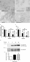
Internalization of holo-Tf and V2R followed by electron microscopy and coimmunoprecipitation. Colocalization of Tf (10-nm gold particles) and V2R-GFP (15-nm gold particles) in LLC-FLAG-V2R cells was shown by gold labeling (A to D). Under basal conditions, Tf and V2R-GFP were located at the plasma membrane both separately and together (A and B, insets). In the presence of Tf for 30 and 60 min, clusters of V2R-GFP were internalized into vesicles of various shapes and sizes (C and D). These images are representative of 3 experiments (bars = 500 nm). Quantification of immunogold-labeled V2R and gold-conjugated Tf was performed (E and F, respectively). Gold-V2R (filled bar) is internalized when cells are warmed to 37°C. This effect is more pronounced in the presence of VP. The number of gold-Tf particles (open bar) is reduced at the plasma membrane at 37°C but is not affected by the presence of VP. The quantification was performed on 6 whole cells (means ± SD). One-way ANOVA was performed between 4°C and other conditions (*P < 0.05 or #P < 0.001). The 37°C control and 37°C + VP conditions are significantly different (*P < 0.05). LLC-V2R-GFP cells expressing TfR1-c-myc were used for an immunoprecipitation assay. V2R-GFP was immunoprecipitated using an anti-GFP polyclonal antibody (G). TfR1-c-myc was detected under basal conditions (lane 2) but not in immunoprecipitations performed in the absence of GFP antibody (lane 1). The TfR1 band intensity increased when cells were exposed to Tf for 30 and 60 min before performing the immunoprecipitation experiment (lanes 3 and 4, respectively). Immunoprecipitated GFP was detected using a monoclonal anti-GFP antibody (G, middle). Quantification of TfR1 band intensity was normalized to the corresponding GFP band intensity (G, bottom). This result is the average of 3 independent experiments (means ± SE, *P < 0.05, 1-way ANOVA).
Fig. 5.
Fig. 5.
Tf-induced V2R internalization is dynamin-dependent. An [3H]-VP binding assay on cells that were or were not infected with dominant-negative Dyn 2 (K44A) adenovirus was performed. The reduction of [3H]-VP binding sites in the presence of holo-Tf (0.3 μM; open bars) was abolished in cells infected with Dyn 2 (K44A) mutant virus. Dyn 2 (K44A) mutant virus did not affect untreated cells (filled bar). This result is representative of 3 independent experiments performed in triplicate (means ± SD; t-test, *P < 0.05, compared with control).
Fig. 6.
Fig. 6.
Tf and VP have a synergistic effect on internalization of plasma membrane V2R-GFP. Western blot analysis of LLC-V2R-GFP cell lysates using an anti-GFP antibody showed that while the mature V2R band intensity was reduced in the presence of VP (filled bars; B), this event was even more pronounced in the presence of both VP and Tf (open bars; B) as shown by quantification of the band intensity representing the mature V2R (A; *P < 0.05, **P < 0.01, 2-way ANOVA, Bonferroni post hoc test). This figure is representative of 5 independent experiments (means ± SE). VP-TMR (seen as yellow spots due to overlap with V2R-GFP) was observed mainly at the plasma membrane of LLC-V2R-GFP cells after 3 min at 37°C (C). VP-TMR-labeled V2R endocytosis is observed 30 min in the absence (D) or in the presence of holo-Tf (E). Quantification of the immunocytochemistry data (F) shows that VP-TMR-labeled V2R endocytosis is slightly increased in the presence of holo-Tf over time. Each time point is the means of 3 different experiments. Each experiment time point has been done in 60 cells (*P < 0.05, 2-way ANOVA, Bonferroni post hoc test; bar = 5 μm).
Fig. 7.
Fig. 7.
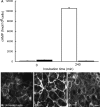
Tf-induced V2R internalization does not trigger the canonical V2R downstream signaling pathway. The presence of Tf for 4 h did not modify the level of intracellular cAMP in LLC-V2R-GFP cells (A; filled bars). In contrast, VP (A; open bars) triggered a significant intracellular cAMP accumulation. The VP/cAMP-sensitive water channel [aquaporin-2 (AQP2)] was located in intracellular vesicles of LLC-PK1 cells under basal conditions (B) or in the presence of Tf (D), whereas VP (10 nM) induced AQP2, a marked plasma membrane accumulation of AQP2 (C). The intracellular cAMP evaluation is the average or representative of 3 experiments (means ± SD). The images are representative of 3 independent experiments (bar = 5 μm).

Similar articles

Cited by

References

    1. Ajioka RS, Kaplan J. Intracellular pools of transferrin receptors result from constitutive internalization of unoccupied receptors. Proc Natl Acad Sci USA 83: 6445–6449, 1986 - PMC - PubMed
    1. Bachvarov DR, Houle S, Bachvarova M, Bouthillier J, Adam A, Marceau F. Bradykinin B(2) receptor endocytosis, recycling, and down-regulation assessed using green fluorescent protein conjugates. J Pharmacol Exp Ther 297: 19–26, 2001 - PubMed
    1. Baldwin GS. Comparison of transferrin sequences from different species. Comp Biochem Physiol B 106: 203–218, 1993 - PubMed
    1. Bider MD, Spiess M. Ligand-induced endocytosis of the asialoglycoprotein receptor: evidence for heterogeneity in subunit oligomerization. FEBS Lett 434: 37–41, 1998 - PubMed
    1. Birnbaumer M. Vasopressin receptor mutations and nephrogenic diabetes insipidus. Arch Med Res 30: 465–474, 1999 - PubMed

Publication types

MeSH terms

LinkOut - more resources