. 2013 Jan 3;493(7430):51-5.
doi: 10.1038/nature11724. Epub 2012 Dec 12.

CCR5 is a receptor for Staphylococcus aureus leukotoxin ED

Affiliations

CCR5 is a receptor for Staphylococcus aureus leukotoxin ED

Francis Alonzo 3rd et al. Nature. .

Abstract

Pore-forming toxins are critical virulence factors for many bacterial pathogens and are central to Staphylococcus aureus-mediated killing of host cells. S. aureus encodes pore-forming bi-component leukotoxins that are toxic towards neutrophils, but also specifically target other immune cells. Despite decades since the first description of staphylococcal leukocidal activity, the host factors responsible for the selectivity of leukotoxins towards different immune cells remain unknown. Here we identify the human immunodeficiency virus (HIV) co-receptor CCR5 as a cellular determinant required for cytotoxic targeting of subsets of myeloid cells and T lymphocytes by the S. aureus leukotoxin ED (LukED). We further demonstrate that LukED-dependent cell killing is blocked by CCR5 receptor antagonists, including the HIV drug maraviroc. Remarkably, CCR5-deficient mice are largely resistant to lethal S. aureus infection, highlighting the importance of CCR5 targeting in S. aureus pathogenesis. Thus, depletion of CCR5(+) leukocytes by LukED suggests a new immune evasion mechanism of S. aureus that can be therapeutically targeted.


Figures
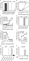
Figure 1
Figure 1. LukED requires CCR5 for cell killing
(a) Viability of cells exposed to different leukotoxins (10 μg/ml). (b) Viability of HUT-R5 cells transduced with control or CCR5 shRNAs. (c) Viability of Jurkat and H9 cells transduced with CCR5 (-R5) or mouse CD24 (-HSA) followed by treatment with LukED. (d) Viability of GHOST cells over-expressing CCR5 and treated with indicated leukotoxins. (e) Viability of HUT-R5 pre-incubated with maraviroc and treated with LukED. (f) Pore-formation, as measured by ethidium bromide uptake, on Jurkat-R5 +/− maraviroc (MVC; 100 ng/ml) followed by incubation with LukED. (g–h) Viability of Jurkat or Jurkat-R5 cells infected with S. aureus (j) in the presence or absence of MVC (k). Means ± standard deviation (n = 3) are shown.
Figure 2
Figure 2. LukE directly interacts with CCR5
(a) Viability of cells treated with α-CCR5 monoclonal antibodies (35 μg/ml) followed by exposure to LukED (10 μg/ml). (b) Membrane association of GFP-LukED (10 μg/ml) to the surface of primary CD4+CCR5+ T cells +/− the indicated monoclonal antibodies (25 μg/ml) as determined by FACS. (c) Membrane association of GFP-LukED (10 μg/ml) on the surface of primary CD4+CCR5+ T cells +/− maraviroc (MVC) (100 ng/ml), CCL5 (5 μg/ml) or on CD4+CCR5 T cells. (d–f) Evaluating the interaction between His-LukE, LukD, or LukED and HA-CCR5 (d) +/− MVC (5 μg/ml) (e), CCL5, CCL4, CCL3 (10 μg/ml) (f), and monoclonal antibodies 45531, 3A9, CXCR4 (35 μg/ml) (f). Immunoblots are representative of at least two independent experiments. (g) Measurement of the interaction of LukE with CCR5 and CXCR4 by SPR. Representative sensorgrams (h) of two experiments performed in duplicate are shown. Where relevant, means ± standard deviation (n = 3) are shown.
Figure 3
Figure 3. LukED kills CCR5+ human memory T cells, macrophages and dendritic cells
(a) Analysis of total CCR5+ primary human T cells (CD3+/CCR5+) incubated with media, LukED (2.5 μg/ml) or maraviroc (MVC; 100 ng/ml) followed by LukED treatment. (b) Susceptibility of T cells isolated from a Δ32-CCR5 or WT-CCR5 donor. Cell viability and CCR5 expression evaluated by flow cytometry as in (a). (c) Measurement of cytokine production of CD4+ T-cells +/− LukED treatment (5 μg/ml) that were stimulated on day 0 with PMA and Ionomycin (P+I; top panel) or cultured in media supplemented with IL-7/IL-15 (20 ng/ml) for 7 days followed by stimulation with P+I (bottom panel). (d) Viability of monocyte-derived macrophages and dendritic cells incubated with LukED (3.0 μg/ml +/− MVC). For FACS plots (a–c) a representative from one of three independent donors is shown. Bar graphs show the mean ± standard deviation of results from three independent donors.
Figure 4
Figure 4. CCR5+ cell killing is important for S. aureus pathogenesis
(a–b) Viability of primary murine peritoneal elicited immune cells from R5+/+ (n=3) or R5−/− (n=3) mice after incubation with LukED (10 μg/ml). (c–d) in vivo viability of recruited immune cells from R5+/+ (n=10) or R5−/− (n=10) mice challenged with live S. aureus Δrot. (e) Bacterial CFU recovered from the kidneys of R5+/+ (n=8) or R5−/− (n=9) mice infected for 96 hours with WT S. aureus. (f) Measurement of serum cytokine and chemokine levels from animals in (e). (g) Quantification of neutrophils and macrophages recovered from infected kidneys 96 hours post-infection. (h) in vivo viability of recruited macrophages from R5+/+ mice challenged with S. aureus WT (n=10) or δlukED (n=10). (i) “Survival” of R5+/+ mice infected with wildtype S. aureus (n=10) or a ΔlukED mutant (n=10) and R5−/− infected with wildtype S. aureus (n=20). FACS plots show a representative from 1 of 10 infected animals. *, p < 0.05; **, p ≤ 0.001; *** p ≤ 0.0001 by 1-way ANOVA (a–b), Student's T test (c–h), and Mantel Cox Test (i). Bar graphs show the mean ± standard deviation.

Comment in

References

    1. Foster TJ. Immune evasion by staphylococci. Nat Rev Microbiol. 2005;3:948–958. - PubMed
    1. Bischofberger M, Iacovache I, Gisou van der Goot F. Pathogenic pore-forming proteins: function and host response. Cell Host Microbe. 2012;12:266–275. doi:10.1016/j.chom.2012.08.005. - PubMed
    1. Menestrina G, et al. Ion channels and bacterial infection: the case of beta-barrel pore-forming protein toxins of Staphylococcus aureus. FEBS Lett. 2003;552:54–60. doi:S0014579303008500 [pii] - PubMed
    1. Dumont AL, et al. Characterization of a new cytotoxin that contributes to Staphylococcus aureus pathogenesis. Mol Microbiol. 2011;79:814–825. doi:10.1111/j.1365-2958.2010.07490.x. - PMC - PubMed
    1. Van de Velde H. Etude sur le mécanisme de la virulence du staphylocoque pyogène. La Cellule. 1894:403–460.

Publication types

MeSH terms