. 2013 Feb;87(4):2287-93.
doi: 10.1128/JVI.02957-12. Epub 2012 Dec 12.

Structure of a dengue virus envelope protein late-stage fusion intermediate

Affiliations

Structure of a dengue virus envelope protein late-stage fusion intermediate

Daryl E Klein et al. J Virol. 2013 Feb.

Abstract

The final stages of dengue virus fusion are thought to occur when the membrane-proximal stem drives the transmembrane anchor of the viral envelope protein (E) toward the fusion loop, buried in the target cell membrane. Crystal structures of E have lacked this essential stem region. We expressed and crystallized soluble mutant forms of the dengue virus envelope protein (sE) that include portions of the juxtamembrane stem. Their structures represent late-stage fusion intermediates. The proximal part of the stem has both intra- and intermolecular interactions, so the chain "zips up" along the trimer seam. The penultimate interaction we detected involves the conserved residue F402, which has hydrophobic contacts with a conserved surface on domain II. These interactions do not require any larger-scale changes in trimer packing. The techniques for expression and crystallization of sE containing stem reported here may allow further characterization of the final stages of flavivirus fusion.


Figures

Fig 1
Fig 1
The dengue virus E protein. (A) Linear representation, showing distribution of the polypeptide chain among three domains I to III, colored red, yellow, and blue, respectively. Domain III leads into a juxtamembrane stem (S) region (green), which links the extracellular region to a carboxy-terminal double-pass transmembrane anchor (TM). (B) Packing of E on the surface of a mature virion. Ninety E dimers are arranged on an icosahedral lattice (10). Symmetry operators are shown in white. Monomers at the center 2-fold axis are outlined with a white line, with a single monomer faded for clarity. (C to F) The fusion process schematized. (C) The dimer lies flat on the viral membrane in the mature prefusion state, with the fusion loops (asterisks) buried in dimer contacts. The two predicted amphipathic helices (green) of the stem lie against the membrane. (D) Low pH causes the dimer to dissociate and the monomers to project outward; the fusion loops are shown buried in the target cell membrane. Domain III folds back and rebinds on the side of domain I, stabilizing a trimer. The arrows indicate that the angle between domains I and II changes, allowing the fusion loops of the three copies of domain II to come together. (E) The stem extends along the seam between two domains II, but the distal stem and membrane anchor are not yet in contact with the fusion loop. (F) All three stems traverse the entire length of domain II, bringing together the TM anchor and the fusion loop, completing membrane merger and pore formation. (G) Amino acid sequence of the stem. Between the two predicted amphipathic helices (H1 and H2) is a conserved region (CON), which varies little across all flaviviruses. The stem sequences of DV1 and DV2 are aligned with those of TBE and West Nile virus (WNV).
Fig 2
Fig 2
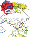
Stem contacts. For clarity, only two monomers of the trimer are shown, with one monomer in surface representation. The orientation is essentially the same as in Fig. 1F. The fusion loop (FL) on the right shows the W101H mutation. The stem is in green. The zoomed inset shows interactions of the stem with the same monomer (color) and the adjacent monomer (gray). Water-bridged hydrogen bonds have been removed. The AB loop of the stem molecule and h-i loop of the adjacent molecule are labeled (designations as in reference 4).
Fig 3
Fig 3
Comparison with other dengue virus E structures. (A) The sE monomer shown is in essentially the same orientation as in Fig. 1F and Fig. 2. DV1 sE is in color. The stemless DV1 trimer (PDB code: 3G7T) is in cyan. The stem and h-i loop are labeled. The most amino-terminal residue of the model (arginine 2) is labeled R2. The principal differences between the two structures are boxed: the AB loop and the fusion loop (FL). (B) The AB loop in the stemless structure participates in a polar cluster linking domains I and III. In the present structure, H317 of the AB loop projects away from the polar cluster. (C) The trimer fusion loops, viewed along the 3-fold axis. The fusion loop W101H mutation and F108 are labeled. Monomers of DV1 sE (PDB code: 3G7T) and DV2 sE (PDB code: 10K8) are superposed on the trimer and shown in cyan and pink, respectively. In the structure described here, F108 adopts the more typical orientation, directed away from the 3-fold axis.
Fig 4
Fig 4
The stem stabilizes a closed trimer conformation. (A) Representation of a hypothetical splayed trimer with the domain I-II hinge in the same orientation as in the DV2 dimer (PDB code: 1OAN). The stem (green) is shown with the intramolecular interactions from Fig. 2: the gap is too great to contact the adjoining molecule. The remainder of the stem, including the disordered residues 404 to 421, is shown as green beads on a string. The angle between 2 fusion loops and the center (C) of the 3-fold axis at the domain I-II junction is 52.3°; the fusion loops are nearly 57 Å apart. The plane of the membrane is shown in light blue bounded by a circle that circumscribes all three fusion loops. The radius (blue rod) (r) defines the distance from the FL to the center of the 3-fold axis (black rod) in the plane of the membrane. (B) In the observed structure, stem contacts bridge between the subunits. The angle is closed by more than 30° compared with the splayed trimer; the fusion loop separation is about 21 Å. The distal part of the stem, including H2, is shown as beads that interact near the fusion loops.

References

    1. Lindenbach BD, Thiel H-J, Rice CM. 2007. Flaviviridae: replication. In Knipe DM, Howley PM, Griffin DE, Lamb RA, Martin MA, Roizman B, Straus SE. (ed), Fields virology, 5th ed, vol 1 Lippincott Williams & Wilkins, Philadelphia, PA
    1. Harrison SC. 2008. Viral membrane fusion. Nat. Struct. Mol. Biol. 15:690–698 - PMC - PubMed
    1. Modis Y, Ogata S, Clements D, Harrison SC. 2003. A ligand-binding pocket in the dengue virus envelope glycoprotein. Proc. Natl. Acad. Sci. U. S. A. 100:6986–6991 - PMC - PubMed
    1. Modis Y, Ogata S, Clements D, Harrison SC. 2004. Structure of the dengue virus envelope protein after membrane fusion. Nature 427:313–319 - PubMed
    1. Allison SL, Schalich J, Stiasny K, Mandl CW, Heinz FX. 2001. Mutational evidence for an internal fusion peptide in flavivirus envelope protein E. J. Virol. 75:4268–4275 - PMC - PubMed

Publication types

Substances

LinkOut - more resources