. 2013 Feb;87(4):2294-306.
doi: 10.1128/JVI.02717-12. Epub 2012 Dec 12.

Outer domain of HIV-1 gp120: antigenic optimization, structural malleability, and crystal structure with antibody VRC-PG04

Affiliations

Outer domain of HIV-1 gp120: antigenic optimization, structural malleability, and crystal structure with antibody VRC-PG04

M Gordon Joyce et al. J Virol. 2013 Feb.

Abstract

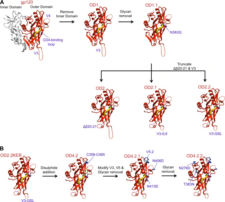
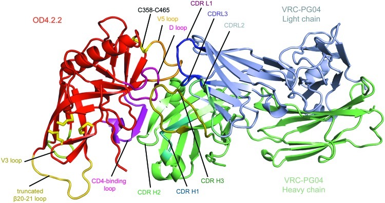
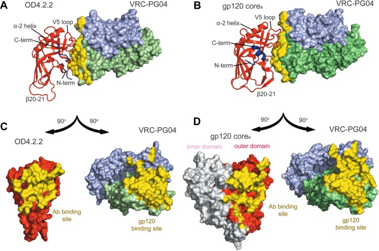
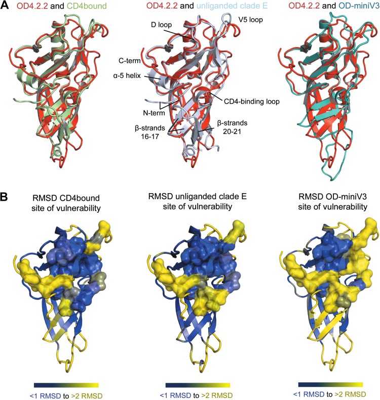
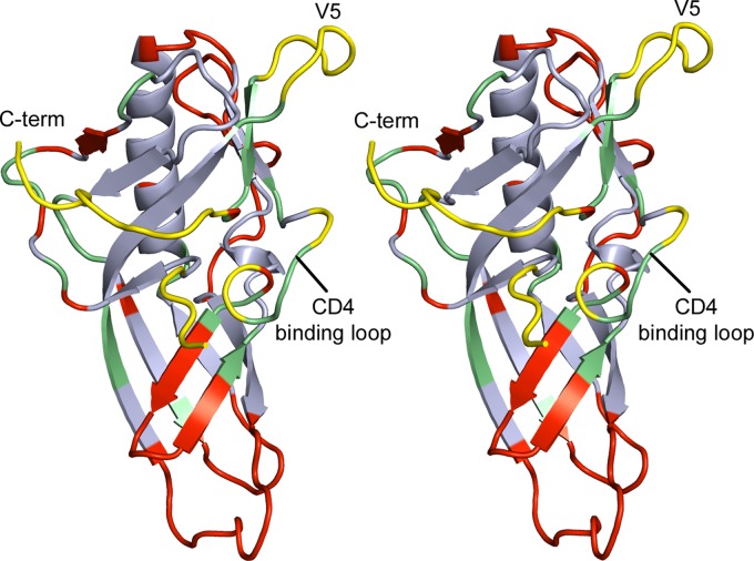
The outer domain of the HIV-1 gp120 envelope glycoprotein contains the epitope for broadly neutralizing antibodies directed to the CD4-binding site, many of which are able to neutralize over 90% of circulating HIV-1 isolates. While the outer domain is conformationally more stable than other portions of the HIV-1 envelope, efforts to express the outer domain as an immunogen for eliciting broadly neutralizing antibodies have not been successful, potentially because natural outer domain variants do not bind strongly to antibodies such as VRC01. In this study, we optimized the antigenic properties of the HIV-1 Env outer domain to generate OD4.2.2, from the KER2018 strain of clade A HIV-1, enabling it to bind antibodies such as VRC01 with nanomolar affinity. The crystal structure of OD4.2.2 in complex with VRC-PG04 was solved at 3.0-Å resolution and compared to known crystal structures including (i) the structure of core gp120 bound by VRC-PG04 and (ii) a circularly permutated version of the outer domain in complex with antibody PGT128. Much of the VRC-PG04 epitope was preserved in the OD4.2.2 structure, though with altered N and C termini conformations. Overall, roughly one-third of the outer domain structure appeared to be fixed in conformation, independent of alterations in termini, clade, or ligand, while other portions of the outer domain displayed substantial structural malleability. The crystal structure of OD4.2.2 with VRC-PG04 provides atomic-level details for an HIV-1 domain recognized by broadly neutralizing antibodies and insights relevant to the rational design of an immunogen that could elicit such antibodies by vaccination.


Figures

Fig 1
Fig 1
Design of HIV-1 gp120 KER2018 clade A OD4.2.2. Sequential rational design based on gp120 structure coupled with binding studies allowed the production of a molecule with high affinity to VRC01-like antibodies. (A) Models of the sequential optimization of the outer domain of HIV-1 gp120 clade B R2 strain shown in red cartoon representation, with the gp120 inner domain shown in gray. Mutations/deletions are indicated by labels and dotted lines. (B) Sequential optimization of HIV-1 clade A KER2018 OD4.2.2 design. Specific mutations are labeled, and the residues are shown in stick representation in blue.
Fig 2
Fig 2
Sequence alignment of outer domain designs. Residues which differ between versions are shown in blue. Residues underlined and in bold were mutated in the outer domain designs. V3-loop and the β20–21 strand residues are in red. Secondary structure elements and names are shown above the sequences with residue numbers also indicated. Predicted glycosylation sites are highlighted in gray, and the four glycosylation sites observed in OD4.2.2 are indicated (formula image) below the OD4.2.2 sequence.
Fig 3
Fig 3
Binding affinity constants of VRC01, VRC-PG04, and IgG b12 to core and outer domain constructs as assessed using Octet Biolayer Interferometry. Sequential truncations of the outer domain molecule resulted in lower binding affinity while structural stabilization, mutagenesis, and glycan removal led to increased affinity for VRC01 and VRC-PG04 antibodies.
Fig 4
Fig 4
Surface plasmon resonance sensorgrams of VRC01, VRC-PG04, and NIH45-46 antibodies immobilized using an anti-Fc antibody and OD4.2.2 flowed over the respective antibodies. The black lines represent the best fit of the kinetic data to a 1:1 binding model. Experiments were carried out at 25°C in PBS buffer (pH 7.4). koff, on-rate constant (M−1s−1); kon, off-rate constant (s−1); KD, equilibrium dissociation constant.
Fig 5
Fig 5
Calorimetry data for the titration of OD4.2.2 with VRC-PG04 antibody in PBS buffer (pH 7.4). Measurements were carried out in triplicate with a representative titration result shown. The top panel shows raw data with the area under each spike proportional to the heat produced at each injection of VRC-PG04 Fab. The lower panel shows integrated areas normalized to the number of moles of VRC-PG04 Fab.
Fig 6
Fig 6
Structure of the outer domain from clade A KER2018, variant OD4.2.2, in complex with the antigen binding domain of antibody VRC-PG04 shown in cartoon representation. Both the light- and heavy-chain regions of VRC-PG04 interact with the outer domain of gp120 in a manner typical of VRC01-like antibodies with the CDR H2 forming strong interactions with the CD4-binding loop. The OD4.2.2 molecule is shown in red except for the V5 loop (orange), D loop (pink), V3 loop (yellow), and truncated β20–21 loop (yellow-orange); disulfide bonds found in the outer domain molecule are shown in yellow stick format. VRC-PG04 is in green and light blue for the heavy and light chains, respectively, except for CDR H1 (teal), CDR H2 (green), CDR H3 (dark green), and CDR L1 (purple), CDR L2 (cyan), and CDR L3 (blue).
Fig 7
Fig 7
Comparison of the OD4.2.2 molecule and clade E coree HIV-1 gp120 molecule in complex with VRC-PG04. Both the gp120 epitopes and antibody paratopes have significant regions of similarity, but additional contacts are made between the heavy chain of VRC-PG04 and OD4.2.2 compared to its binding site observed in the HIV-1 coree complex structure, while VRC-PG04 light-chain contacts are reduced. (A) OD4.2.2 (cartoon format with the N and C termini regions in blue) in complex with VRC-PG04 (surface representation). The buried surface area for VRC-PG04 is shown in yellow. (B) The outer domain of HIV-1 gp120 coree (cartoon format; the inner domain is omitted for clarity) in complex with VRC-PG04 (surface representation) is shown with the VRC-PG04 buried surface area in yellow. (C) OD4.2.2 and VRC-PG04 are rotated 90° in opposite directions, respectively, to allow visualization of the buried surface area between the epitope of the gp120 outer domain and the paratope of the VRC-PG04 antibody with the buried interactive surfaces shown in yellow. (D) HIV-1 gp120 coree molecule and the VRC-PG04 antibody are rotated to allow visualization of their respective epitopes and paratopes which are shown in yellow surface representation.
Fig 8
Fig 8
Structural comparison of OD4.2.2 to HIV-1 gp120 in the CD4-bound form as observed in the CD4-liganded complex and unliganded clade E coree and the HXBc2 circularly permutated OD-miniV3. Comparisons indicate that there are significant differences in the HIV-1 site of vulnerability, the N- and C-terminal region of the outer domain, and regions adjacent to β-strands 16 and 17. (A) Structural overlay of OD4.2.2 (red) with the gp120 outer domain of CD4-bound gp120 (green) and unliganded clade E gp120 (light blue) and the circularly permutated HXBc2 OD-miniV3 outer domain (teal). (B) OD4.2.2 is shown in cartoon representation colored according to RMSD comparisons with other outer domains of gp120 (blue, <1 Å RMSD; yellow, >2 Å RMSD); the previously defined site of vulnerability is shown in semitransparent surface representation.
Fig 9
Fig 9
Sequence alignment of outer domain molecules (OD4.2.2 and OD-miniV3) and the outer domains of the CD4-bound HXBc2 molecule and unbound clade E molecule. Residues highlighted in red form the site of vulnerability of the CD4-receptor binding site.
Fig 10
Fig 10
Stereo view of the OD4.2.2 molecule. Analysis and comparisons of this structure indicate regions which are conserved between outer domain molecules regardless of sequence or design (light blue), regions which are in the CD4-bound conformation (green), areas which are dissimilar across the two outer domain molecules (red), and regions which should be targeted for further optimization/stabilization (yellow) in order to produce an optimal outer domain immunogen.

References

    1. Korber B, Gaschen B, Yusim K, Thakallapally R, Kesmir C, Detours V. 2001. Evolutionary and immunological implications of contemporary HIV-1 variation. Br. Med. Bull. 58:19–42 - PubMed
    1. Kwong PD, Doyle ML, Casper DJ, Cicala C, Leavitt SA, Majeed S, Steenbeke TD, Venturi M, Chaiken I, Fung M, Katinger H, Parren PW, Robinson J, Van Ryk D, Wang L, Burton DR, Freire E, Wyatt R, Sodroski J, Hendrickson WA, Arthos J. 2002. HIV-1 evades antibody-mediated neutralization through conformational masking of receptor-binding sites. Nature 420:678–682 - PubMed
    1. Wei X, Decker JM, Wang S, Hui H, Kappes JC, Wu X, Salazar-Gonzalez JF, Salazar MG, Kilby JM, Saag MS, Komarova NL, Nowak MA, Hahn BH, Kwong PD, Shaw GM. 2003. Antibody neutralization and escape by HIV-1. Nature 422:307–312 - PubMed
    1. Zhou T, Georgiev I, Wu X, Yang ZY, Dai K, Finzi A, Kwon YD, Scheid JF, Shi W, Xu L, Yang Y, Zhu J, Nussenzweig MC, Sodroski J, Shapiro L, Nabel GJ, Mascola JR, Kwong PD. 2010. Structural basis for broad and potent neutralization of HIV-1 by antibody VRC01. Science 329:811–817 - PMC - PubMed
    1. Wu X, Zhou T, Zhu J, Zhang B, Georgiev I, Wang C, Chen X, Longo NS, Louder M, McKee K, O'Dell S, Perfetto S, Schmidt SD, Shi W, Wu L, Yang Y, Yang ZY, Yang Z, Zhang Z, Bonsignori M, Crump JA, Kapiga SH, Sam NE, Haynes BF, Simek M, Burton DR, Koff WC, Doria-Rose NA, Connors M, Mullikin JC, Nabel GJ, Roederer M, Shapiro L, Kwong PD, Mascola JR. 2011. Focused evolution of HIV-1 neutralizing antibodies revealed by structures and deep sequencing. Science 333:1593–1602 - PMC - PubMed

Publication types

MeSH terms