. 2012 Dec 26;109(52):21480-5.
doi: 10.1073/pnas.1216238110. Epub 2012 Dec 10.

Structural basis of toxicity and immunity in contact-dependent growth inhibition (CDI) systems

Affiliations

Structural basis of toxicity and immunity in contact-dependent growth inhibition (CDI) systems

Robert P Morse et al. Proc Natl Acad Sci U S A. .

Abstract

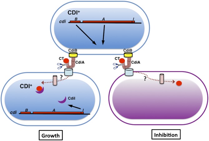
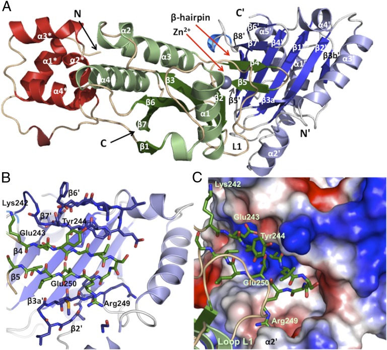
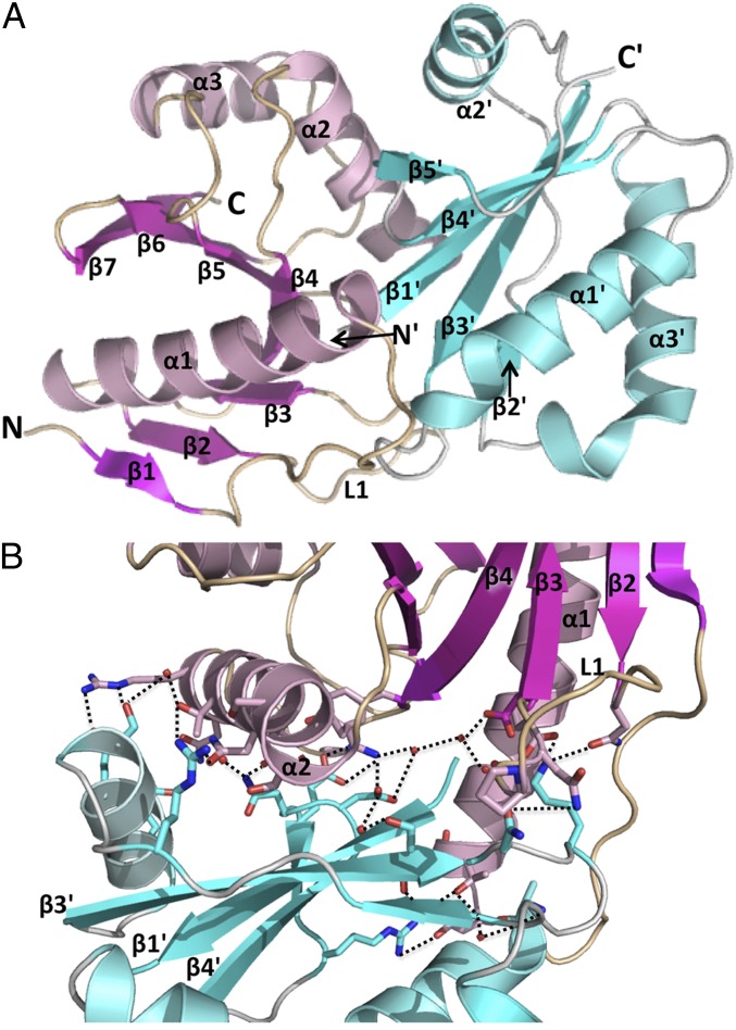
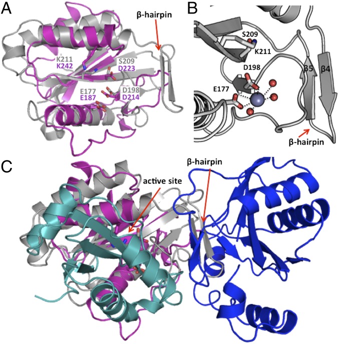
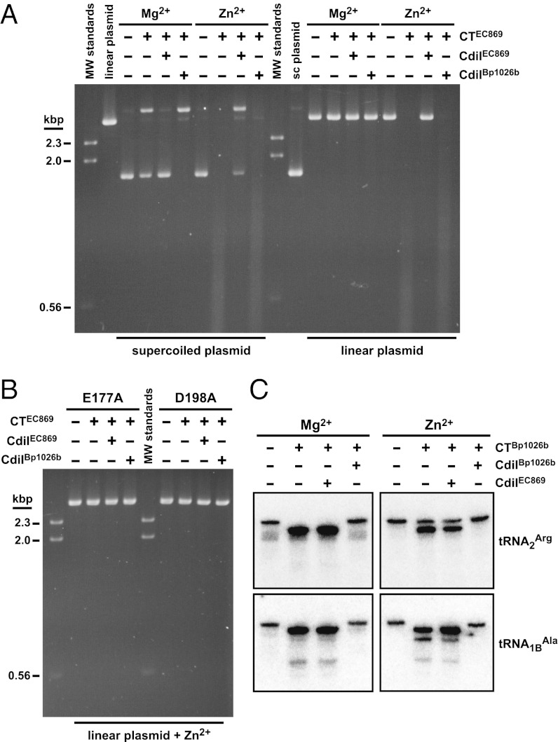
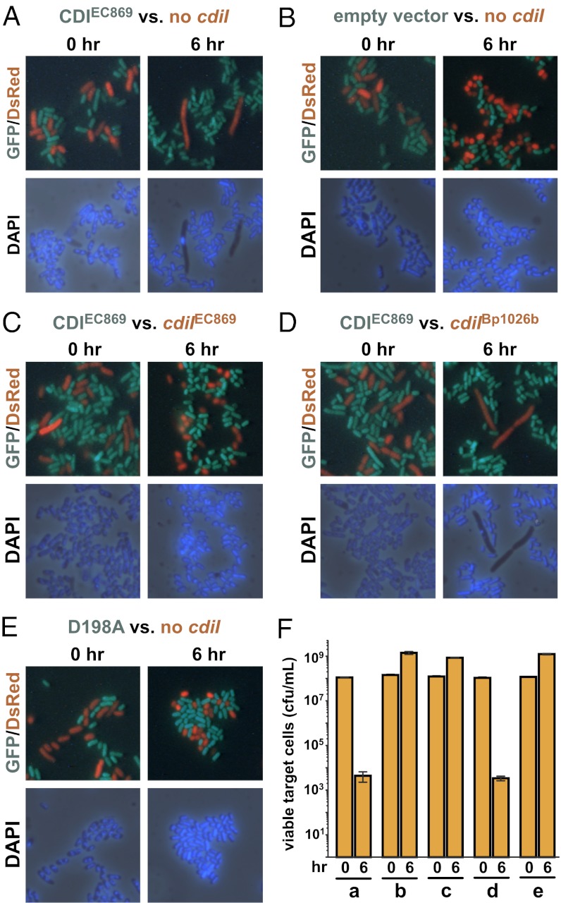
Contact-dependent growth inhibition (CDI) systems encode polymorphic toxin/immunity proteins that mediate competition between neighboring bacterial cells. We present crystal structures of CDI toxin/immunity complexes from Escherichia coli EC869 and Burkholderia pseudomallei 1026b. Despite sharing little sequence identity, the toxin domains are structurally similar and have homology to endonucleases. The EC869 toxin is a Zn(2+)-dependent DNase capable of completely degrading the genomes of target cells, whereas the Bp1026b toxin cleaves the aminoacyl acceptor stems of tRNA molecules. Each immunity protein binds and inactivates its cognate toxin in a unique manner. The EC869 toxin/immunity complex is stabilized through an unusual β-augmentation interaction. In contrast, the Bp1026b immunity protein exploits shape and charge complementarity to occlude the toxin active site. These structures represent the initial glimpse into the CDI toxin/immunity network, illustrating how sequence-diverse toxins adopt convergent folds yet retain distinct binding interactions with cognate immunity proteins. Moreover, we present visual demonstration of CDI toxin delivery into a target cell.


Conflict of interest statement

The authors declare no conflict of interest.

Figures

Fig. 1.
Fig. 1.
The CDI pathway. CDI+ cells containing the cdiBAI gene cluster express CdiB and CdiA at the cell surface. Contact between CdiA and the BamA receptor on the surface of target cells results in delivery of the CdiA-CT toxin into the target cell. The mechanisms of toxin translocation are not understood, but BamA (4) and unknown inner membrane components are hypothesized to mediate transport. Cells carrying the identical CDI system (depicted in blue cells) are protected from growth inhibition by the CdiI immunity protein, which specifically binds and inactivates the CdiA-CT toxin. Nonimmune cells are inhibited by CdiA-CT (depicted in purple cells).
Fig. 2.
Fig. 2.
Structure of the EC869 CdiA-CTo11/CdiIo11 complex. (A) Ribbon representation of the CdiA-CTo11EC869/CdiI o11EC869 complex. CdiA-CTo11EC869 contains two domains, an N-terminal α-helical bundle (red) and a C-terminal α/β nuclease domain (green). The four helices marked with asterisks (*) form the N-terminal helical bundle of CdiA-CTo11EC869. The CdiIo11EC869 immunity protein (blue) is composed of a single α/β domain. The secondary structure elements of each protein are identified and their N and C termini are indicated. All immunity protein elements are denoted with a prime symbol () to differentiate them from the toxin secondary structure elements. The active site Zn2+ ion is depicted as a purple sphere. (B) The CdiA-CTo11EC869 and CdiIo11EC869 proteins interact through β-augmentation. The β4,β5-hairpin of CdiA-CTo11EC869 (carbon atoms, green) inserts into the CdiI o11EC869 immunity protein (carbon atoms, blue) to form a six-stranded antiparallel β-sheet. β-hairpin residues and ion pairs are represented as sticks (where the oxygen, nitrogen, and sulfur atoms are colored red, dark blue, and yellow, respectively). (C) CdiA-CTo11EC869 β-hairpin (green sticks) along with the extended loop region L1 fits snugly into the molecular surface representation of CdiI o11EC869. White surfaces represent hydrophobic regions, and the red and blue surfaces indicate negative and positive electrostatic potential, respectively.
Fig. 3.
Fig. 3.
Structure of the Bp1026b CdiA-CTII/CdiIII complex. (A) The CdiA-CTllBp1026b toxin (pink) and CdiIllBp1026b immunity protein (cyan) are depicted in ribbon representation with secondary structure elements indicated. All immunity protein elements are denoted with a prime symbol () to differentiate them from the toxin secondary structure elements. (B) The interface between CdiA-CTllBp1026b (pink) and CdiIllBp1026b (cyan) is formed by an extensive network of ion pairs and hydrogen bonds. Within the network, interacting residue side chains are represented as sticks (oxygen and nitrogen atoms are colored red and blue, respectively), water molecules as red spheres, and interacting bonds as black dotted lines.
Fig. 4.
Fig. 4.
Structural superimposition of EC869 and Bp1026b CdiA-CT/CdiI protein complexes. (A) Predicted active site residues of the EC869 and Bp1026b toxin domains. The two toxin domains are superimposed and active site residues are rendered as stick representations. EC869 and Bp1026b carbon atoms are colored gray and pink, respectively; oxygen and nitrogen atoms are colored red and blue, respectively. (B) Coordination of Zn2+ within the CdiA-CTo11EC869 active site. The Zn2+ ion is depicted as a purple sphere, ordered waters as smaller red spheres, and interacting bonds with Zn2+ are depicted as black dotted lines. (C) Superimposition of the EC869 and Bp1026b CdiA-CT/CdiI protein complexes. Ribbon representations of CdiA-CTllBp1026b and CdiIllBp1026b are colored pink and cyan, respectively, and the C-terminal domains of CdiA-CTo11EC869 and CdiI o11EC869 are colored gray and blue, respectively. The C-terminal toxin domains superimpose upon one another, whereas the immunity proteins do not. The N-terminal α-helical domain of CdiA-CTo11EC869 has been omitted for clarity.
Fig. 5.
Fig. 5.
CdiA-CT toxins have distinct nuclease activities. (A) DNase activity of the CdiA-CTo11EC869 toxin on supercoiled and linear plasmid substrates. Plasmid DNA was incubated with purified CdiA-CTo11EC869 in the presence of either Mg2+ or Zn2+ and reactions were analyzed by agarose gel electrophoresis and ethidium bromide staining. Reactions also included either purified CdiIo11EC869 or CdiIIIBp1026b immunity proteins where indicated. Untreated supercoiled linear plasmid substrates were included as controls for the migration of undigested DNA. The migration positions of linear molecular weight (MW) DNA standards are indicated in kilobase pairs (kbp). (B) Mutation of predicted active site residues ablates CdiA-CTo11EC869 DNase activity. Linear plasmid DNA was incubated with purified CdiA-CTo11EC869 containing the Glu177Ala (E177A) and Asp198Ala (D198A) mutations in buffer supplemented with Zn2+. Reactions also contained CdiIo11EC869 or CdiIIIBp1026b immunity proteins where indicated. (C) The CdiA-CTIIBp1026b toxin has tRNase activity. Purified E. coli tRNA was treated with CdiA-CTIIBp1026b toxin in reactions supplemented with Mg2+ or Zn2+. Reactions contained CdiIo11EC869 or CdiIIIBp1026b immunity proteins where indicated and were run on denaturing polyacrylamide gels and analyzed by Northern blot hybridization using radiolabeled probes to tRNA2Arg and tRNA1BAla.
Fig. 6.
Fig. 6.
The CdiA-CTo11EC869 toxin degrades DNA during contact-dependent growth inhibition (CDI). GFP-labeled E. coli inhibitor cells (green) were mixed with DsRed-labeled target cells (red) and grown in shaking broth cultures. Cocultures were sampled at 0 and 6 h and stained with DAPI to visualize cellular DNA by fluorescence microscopy. (A) EC93-EC869o11 inhibitor cells versus targets that lack an immunity gene. (B) Mock inhibitor cells (carrying an empty vector cosmid) versus targets that lack an immunity gene. (C) EC93-EC869o11 inhibitors versus target cells that carry the cognate cdiIo11EC869 gene. (D) EC93-EC869o11 inhibitors versus target cells that carry the noncognate cdiIIIBp1026b immunity gene. (E) EC93-EC869o11 inhibitors carrying the Asp198Ala (D198A) missense mutation versus target cells that lack an immunity gene. (F) Quantification of viable target cells during CDI. The number of viable target cells at 0 and 6 h were determined as colony forming units (cfu) per milliliter. The data from competitions corresponding to panels AE are indicated. Values are the mean ± SEM for three independent experiments.

References

    1. Aoki SK, et al. A widespread family of polymorphic contact-dependent toxin delivery systems in bacteria. Nature. 2010;468(7322):439–442. - PMC - PubMed
    1. Aoki SK, et al. Contact-dependent inhibition of growth in Escherichia coli. Science. 2005;309(5738):1245–1248. - PubMed
    1. Kajava AV, et al. Beta-helix model for the filamentous haemagglutinin adhesin of Bordetella pertussis and related bacterial secretory proteins. Mol Microbiol. 2001;42(2):279–292. - PubMed
    1. Aoki SK, et al. Contact-dependent growth inhibition requires the essential outer membrane protein BamA (YaeT) as the receptor and the inner membrane transport protein AcrB. Mol Microbiol. 2008;70(2):323–340. - PMC - PubMed
    1. Nikolakakis K, et al. The toxin/immunity network of Burkholderia pseudomallei contact-dependent growth inhibition (CDI) systems. Mol Microbiol. 2012;84(3):516–529. - PMC - PubMed

Publication types

MeSH terms

LinkOut - more resources