. 2012 Dec 26;109(52):21438-43.
doi: 10.1073/pnas.1217409110. Epub 2012 Dec 10.

Distinct mechanisms mediate naive and memory CD8 T-cell tolerance

Affiliations

Distinct mechanisms mediate naive and memory CD8 T-cell tolerance

Evan R Jellison et al. Proc Natl Acad Sci U S A. .

Abstract

Peripheral tolerance to developmentally regulated antigens is necessary to sustain tissue homeostasis. We have now devised an inducible and reversible system that allows interrogation of T-cell tolerance induction in endogenous naïve and memory CD8 T cells. Our data show that peripheral CD8 T-cell tolerance can be preserved through two distinct mechanisms, antigen addiction leading to anergy for naïve T cells and ignorance for memory T cells. Induction of antigen in dendritic cells resulted in substantial expansion and maintenance of endogenous antigen-specific CD8 T cells. The self-reactive cells initially exhibited effector activity but eventually became unresponsive. Upon antigen removal, the antigen-specific population waned, resulting in development of a self-specific memory subset that recalled to subsequent challenge. In striking contrast to naïve CD8 T cells, preexisting antigen-specific memory CD8 T cells failed to expand after antigen induction and essentially ignored the antigen despite widespread expression by dendritic cells. The inclusion of inflammatory signals partially overcame memory CD8 T-cell ignorance of self-antigen. Thus, peripheral CD8 T-cell tolerance for naïve CD8 T cells depended on the continuous presence of antigen, whereas memory CD8 T cells were prohibited from autoreactivity in the absence of inflammation.


Conflict of interest statement

The authors declare no conflict of interest.

Figures

Fig. 1.
Fig. 1.
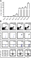
Antigen expression is tightly controlled in triple transgenic mice. (A) CFSE-labeled Rag−/− OT-I cells (5 × 10⁵) were transferred to the indicated mice 1 d before administration of 10 μg/mL doxycycline in the drinking water. Six days later, donor OT-I splenocytes were examined for CFSE loss by flow cytometry. Values indicate the percentage of OT-I cells among CD8+ T cells in the spleen. (B) C57BL/6 or CST BMC mice were infected i.v. with 1 × 10⁵ pfu VSV-SED. Splenocytes were stained with the indicated MHC I tetramers or antibodies and analyzed by flow cytometry 7 d after infection. This experiment was performed three times with 3–4 mice per group.
Fig. 2.
Fig. 2.
Expansion and activation of endogenous T cells in response to induced self-antigen. CST BMC mice were treated with doxycycline, and the antigen-specific CD8 T-cell response was monitored. (A) Number of OVA257-Kb specific T cells over the course of antigen induction. (B) Spleen, peripheral lymph nodes, and mesenteric lymph nodes were pooled and stained with OVA257Kb tetramer and anti-CD8 antibody and enriched by magnetic sorting. Enriched cells were further stained with the indicated antibodies and analyzed by flow cytometry.
Fig. 3.
Fig. 3.
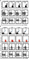
Analysis of self-specific CD8 T cells in lymphoid and nonlymphoid tissues. (A) Lymphocytes from secondary lymphoid organs were stained as in Fig. 2A, but without magnetic sorting. (B) Lymphocytes from CST BMC mice were isolated from indicated organs after 35 d of doxycycline treatment, stained as indicated, and analyzed by flow cytometry. These experiments were performed at least two times with a minimum of three mice per group.
Fig. 4.
Fig. 4.
Maintenance of self-specific CD8 T cells and the generation of memory T cells by self antigen. (A) OT-I cells were transferred to CST BMC mice that had been treated with doxycycline for 45 d. The mice were given BrdU for the next week, and BrdU incorporation was determined in both endogenous and donor OT-I populations. These experiments were performed at least two times with a minimum of three mice per group. (BE) Antigen was induced for 10 d in CST BMC mice and then doxycycline was withdrawn, and the response was monitored in the blood at the indicated times for the indicated parameters. SLEC, CD127- KLRG1+ short-lived effector cells; EEC, KLRG CD127 early effector cells; MPEC, CD127+ KLRG1 memory precursor effector cell; DPEC, KLRG1+ CD127+ double positive effector cells. (F) Cells from secondary lymphoid organs were enriched for tetramer positive cells and were analyzed by flow cytometry 35 d after doxycycline withdrawal. (G) Naïve CST BMC mice or mice treated with doxycycline for 10 d followed by 35 d without doxycyline were infected with VSV-SED. Graph represents the percent of tetramer+ CD8+ T cells in blood. These experiments were performed three times with a minimum of three mice per group.
Fig. 5.
Fig. 5.
Pathogen-specific memory cells ignore self-antigen. (A) Cells from secondary lymphoid organs of CST BMC mice treated with doxycycline and BrdU for 7 d were stained and enriched for tetramer binding cells. (BE), Tetramer+ cells specific for either OVA or VSV nucleoprotein (N) were analyzed from CST BMC mice infected with 1 × 10⁵ pfu VSV-SED. Sixty days after infection, mice were given BrdU and additionally treated as indicated with nothing or with doxycycline (B), or infected i.p. with 1 × 10⁶ pfu recombinant vaccinia virus expressing either OVA (C) or VSV N (D). Seven days later, tetramer+ cells in the spleen were quantitated and analyzed for BrdU incorporation. Values represent the means derived from at least three mice ± SEM. This experiment was performed three times with a minimum of three mice per group.

References

    1. Gallegos AM, Bevan MJ. Central tolerance: Good but imperfect. Immunol Rev. 2006;209:290–296. - PubMed
    1. Xing Y, Hogquist KA. T-cell tolerance: Central and peripheral. Cold Spring Harb Perspect Biol. 2012;4(6):4. - PMC - PubMed
    1. Metzger TC, Anderson MS. Control of central and peripheral tolerance by Aire. Immunol Rev. 2011;241(1):89–103. - PMC - PubMed
    1. Liston A, Rudensky AY. Thymic development and peripheral homeostasis of regulatory T cells. Curr Opin Immunol. 2007;19(2):176–185. - PubMed
    1. Pape KA, Merica R, Mondino A, Khoruts A, Jenkins MK. Direct evidence that functionally impaired CD4+ T cells persist in vivo following induction of peripheral tolerance. J Immunol. 1998;160(10):4719–4729. - PubMed

Publication types

LinkOut - more resources