. 2012 Dec 26;109(52):21301-6.
doi: 10.1073/pnas.1213818110. Epub 2012 Dec 11.

Versatile design and synthesis platform for visualizing genomes with Oligopaint FISH probes

Affiliations

Versatile design and synthesis platform for visualizing genomes with Oligopaint FISH probes

Brian J Beliveau et al. Proc Natl Acad Sci U S A. .

Abstract

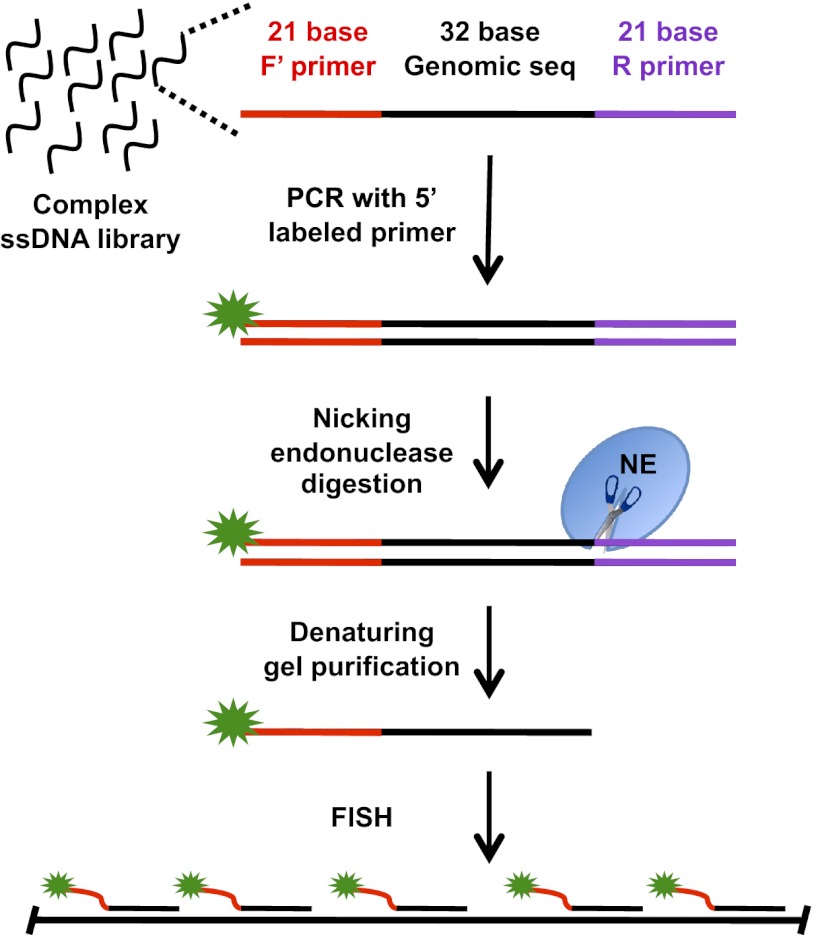
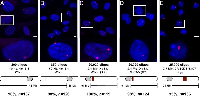
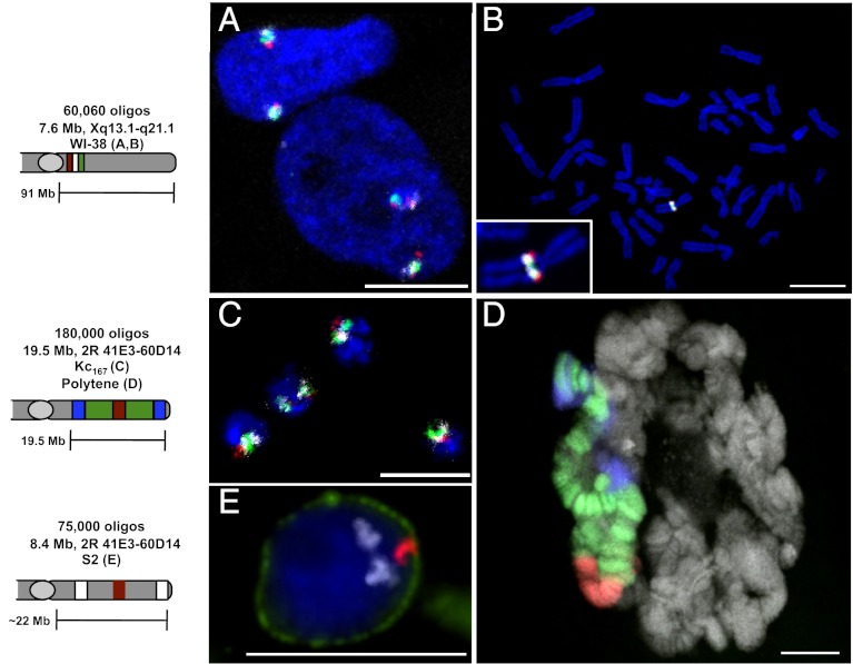
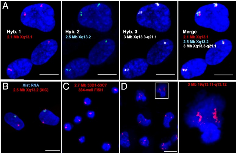
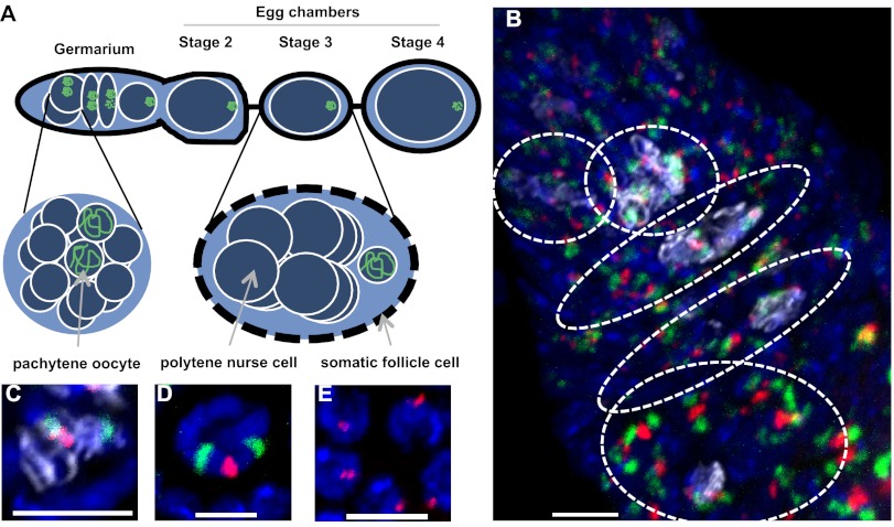
A host of observations demonstrating the relationship between nuclear architecture and processes such as gene expression have led to a number of new technologies for interrogating chromosome positioning. Whereas some of these technologies reconstruct intermolecular interactions, others have enhanced our ability to visualize chromosomes in situ. Here, we describe an oligonucleotide- and PCR-based strategy for fluorescence in situ hybridization (FISH) and a bioinformatic platform that enables this technology to be extended to any organism whose genome has been sequenced. The oligonucleotide probes are renewable, highly efficient, and able to robustly label chromosomes in cell culture, fixed tissues, and metaphase spreads. Our method gives researchers precise control over the sequences they target and allows for single and multicolor imaging of regions ranging from tens of kilobases to megabases with the same basic protocol. We anticipate this technology will lead to an enhanced ability to visualize interphase and metaphase chromosomes.


Conflict of interest statement

The authors declare no conflict of interest.

Figures

Fig. 1.
Fig. 1.
Each oligonucleotide in the library is composed of 32 bases of genomic sequence flanked by 21-base primer sequences. One of the primers carries a 5′ fluorophore, whereas the other contains a recognition site for a nicking endonuclease (NE) (37). A nicking reaction followed by denaturing gel electrophoresis yields 53-base ssDNAs.
Fig. 2.
Fig. 2.
Oligopaints efficiently label interphase human and Drosophila nuclei. (A) Probe set of 200 oligos targeting 10 kb at human 4p16.1 was hybridized to WI-38 (2N) cells. (B) Probe set targeting the region shown in A, but extended to 850 oligos targeting 52 kb was hybridized to WI-38 cells. (C and D) Probe set of 20,020 oligos targeting 2.1 Mb at human Xq13.1 was hybridized to WI-38 cells (XX) (C) or MRC-5 (XY 2N) cells (D). (E) Probe set of 25,000 oligos targeting 2.7 Mb at 50D1-53C7 on Drosophila 2R was hybridized to Kc167 (4N) cells. Enlarged image of the Inset is shown beneath each micrograph. (Bottom) Labeling efficiencies presented as the percentage of cells that displayed at least one FISH focus (SI Appendix, Table S2). All probe sets were labeled with TYE563 (Cy3 mimic; red); DNA was identified with DAPI (blue). (Scale bars, 5 µM.) Images are maximum Z projections. Each micrograph was acquired using parameters optimized for entire fields of cells; thus, the sizes of the foci do not necessarily correlate with the sizes of the targeted regions.
Fig. 3.
Fig. 3.
Multicolor FISH with Oligopaints. (A and B) Three 20,020 oligo probe sets targeting adjacent regions at human Xq13.1, Xq13.2, and Xq13.3-q21.1 (SI Appendix, Fig. S1A and Table S3) were used to produce 3-color FISH images from WI-38 (XX) interphase (A) and primary metaphase (Abbott Molecular) (B) chromosomes. Probe sets were labeled with TYE563 (Cy3 mimic; red), TYE665 (Cy5 mimic; white), or 6-FAM (green), respectively. (C and D) Probe set of 180,000 oligos was used to paint a multicolor banding pattern from 41E3 to 60D14 (SI Appendix, Fig. S1B and Table S3) on Drosophila 2R in interphase Kc167 (4N) nuclei (C) and salivary gland polytene chromosomes (D) with the following pattern: 41E3–44C4 [white (C) or blue (D); 25,000 TYE665-labeled oligos], 44C4–50C9 (green; 52,500 6-FAM–labeled oligos), 50D1–53C7 (red; 25,000 TYE563-labeled oligos), 53C9–58B6 (green, 52,500 6-FAM–labeled oligos), and 58D2–60D14 [white (C) or blue (D); 25,000 TYE665-labeled oligos]. (E) Two probe sets were combined to span Drosophila 2R. One probe set was composed of 25,000 TYE563 (red)-labeled oligos targeting 50D1–53C7, whereas the second was composed of 25,000 TYE665 (white)-labeled oligos targeting 41E3–44C4 and 25,000 TYE665 (white)-labeled oligos targeting 58D2–60D14. The nuclear envelope was stained with wheat germ agglutinin conjugated to Alexa Fluor 488 (green). A, C, and E are maximum Z projections, whereas B and D are single Z slices. DNA was identified with DAPI (blue for AC and E; gray for D). (Scale bars, 10 µM.)
Fig. 4.
Fig. 4.
Multiple applications for Oligopaints. (A) Repeated rounds of hybridization in WI-38 (XX 2N) cells using a probe set composed of 20,020 TYE563 (Cy3 mimic; red)-labeled oligos targeting 2.1 Mb at Xq13.1, a probe set composed of 20,020 TYE665 (Cy5 mimic; green)-labeled oligos targeting 2.5 Mb at Xq13.2, and a probe set composed of 20,020 6-FAM (white)-labeled oligos targeting 3.0 Mb at Xq13.3–q21.1. Once a slide was hybridized with a probe set labeled with a given fluorophore (e.g., Tye563 for Hyb 1), the slide was then scanned for the presence of that fluorophore after all successive hybridizations. For all panels, DNA was identified by DAPI (blue). Also see SI Appendix, Fig. S9. (B) Simultaneous RNA/DNA FISH using a probe set composed of 20,020 TYE563 (red)-labeled oligos targeting the XIC at Xq13.2 and spanning 2.5 Mb and a probe set composed of 96 6-FAM (green)-labeled oligos targeting the Xist RNA in WI-38 cells (42). Also see SI Appendix, Fig. S10. (C) Automated imaging of a probe set composed of 25,000 TYE563-labeled oligos targeting a 2.7-Mb region at 50D1–53C7 on Drosophila 2R in Kc167 cells (4N; single focus reflects pairing of homologous chromosomes in Drosophila) seeded in a 384-well plate. Also see SI Appendix, Fig. S11. (D) Examples of morphologies produced by a probe set composed of 20,020 Cy3-labeled oligos targeting 3 Mb at human chr19q13.11–q13.12 in WI-38 cells. (Right) Enlarged image of Inset. All images are maximum Z projections. (Scale bars, 10 µM.)
Fig. 5.
Fig. 5.
Oligopaints efficiently label nuclei from whole-mounted Drosophila ovaries. (A) Cartoon of a Drosophila ovariole displaying three cell types: the pachytene oocytes and polytene nurse cells within the meiotic cysts and the somatic follicle cells that encase them. (B) Drosophila germarium labeled with two probe sets composed of 25,000 TYE563 (Cy3 mimic; red)-labeled oligos targeting a 2.7-Mb region at 50D1–53C7 and an additional set composed of two pools of 25,000 TYE665 (Cy5 mimic; green)-labeled oligos targeting 41E3–44C4 and 58D2–60D14 (green), all regions located on Drosophila 2R. An antibody to the synaptonemal complex component C(3)G (white) was used to identify oocytes. Hashed circles demarcate the meiotic cysts. (C and D) Same probe set as described for B in a magnified view of a single oocyte (C) and a polytene nurse cell (D). (E) The 50D1–53C7 probe set in a magnified view of follicle cells. (Scale bars, 5 µM.) For all panels, DNA was identified by DAPI (blue).

Similar articles

Cited by

References

    1. Dekker J, Rippe K, Dekker M, Kleckner N. Capturing chromosome conformation. Science. 2002;295(5558):1306–1311. - PubMed
    1. Lieberman-Aiden E, et al. Comprehensive mapping of long-range interactions reveals folding principles of the human genome. Science. 2009;326(5950):289–293. - PMC - PubMed
    1. de Wit E, de Laat W. A decade of 3C technologies: Insights into nuclear organization. Genes Dev. 2012;26(1):11–24. - PMC - PubMed
    1. Tanizawa H, Noma K. Unravelling global genome organization by 3C-seq. Semin Cell Dev Biol. 2012;23(2):213–221. - PMC - PubMed
    1. Pardue ML, Gall JG. Molecular hybridization of radioactive DNA to the DNA of cytological preparations. Proc Natl Acad Sci USA. 1969;64(2):600–604. - PMC - PubMed

Publication types

Substances