. 2014 Apr;24(4):968-77.
doi: 10.1093/cercor/bhs380. Epub 2012 Dec 12.

Role of the prefrontal cortex in altered hippocampal-accumbens synaptic plasticity in a developmental animal model of schizophrenia

Affiliations

Role of the prefrontal cortex in altered hippocampal-accumbens synaptic plasticity in a developmental animal model of schizophrenia

Pauline Belujon et al. Cereb Cortex. 2014 Apr.

Abstract

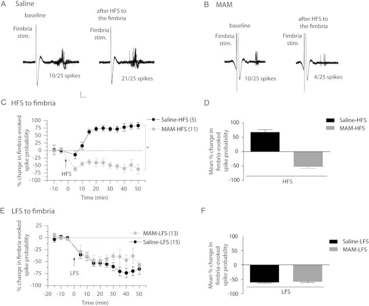
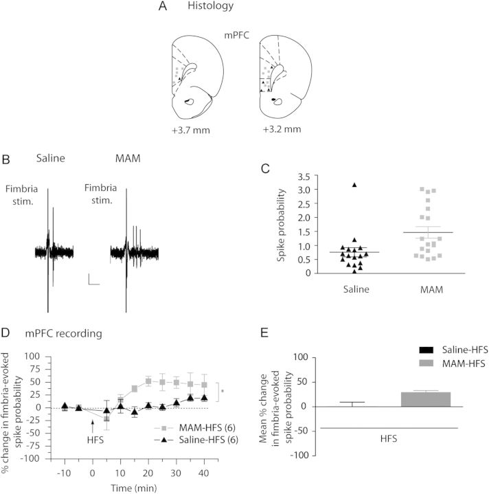
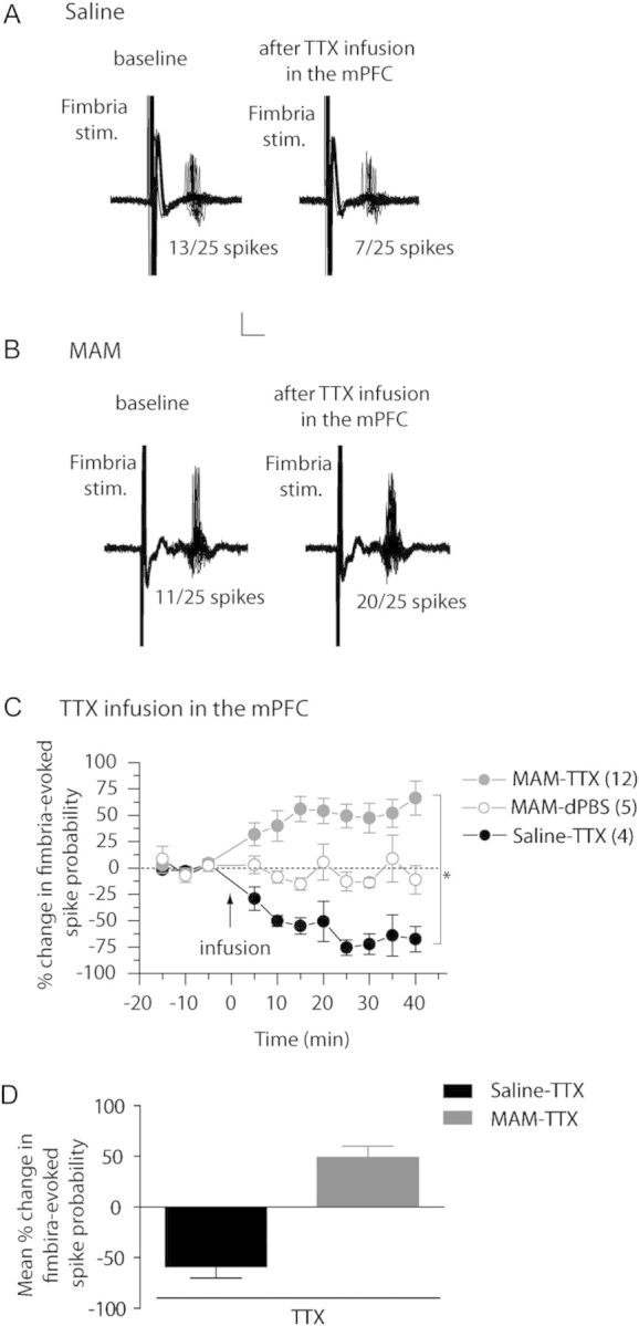
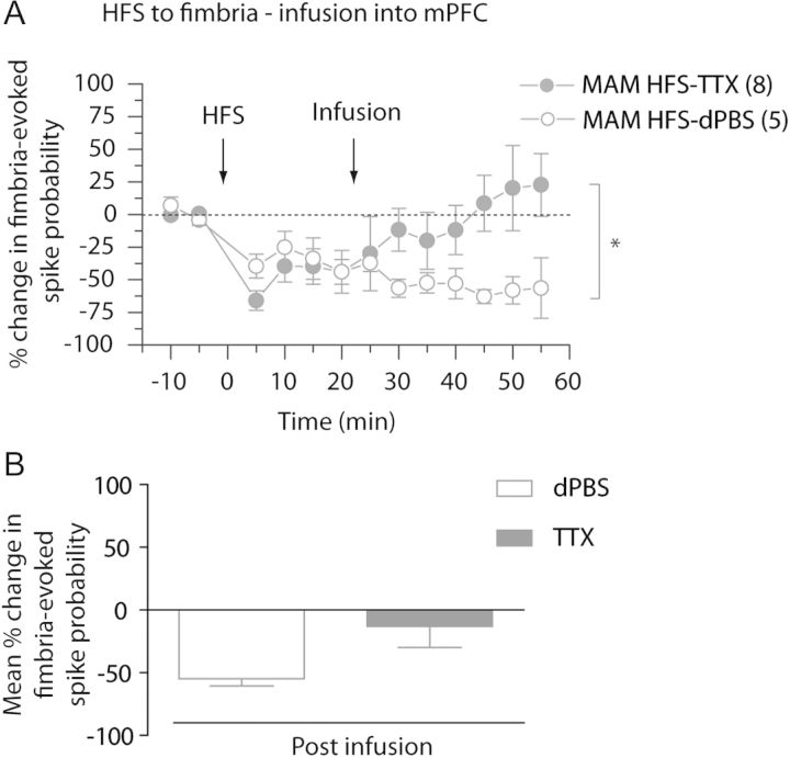
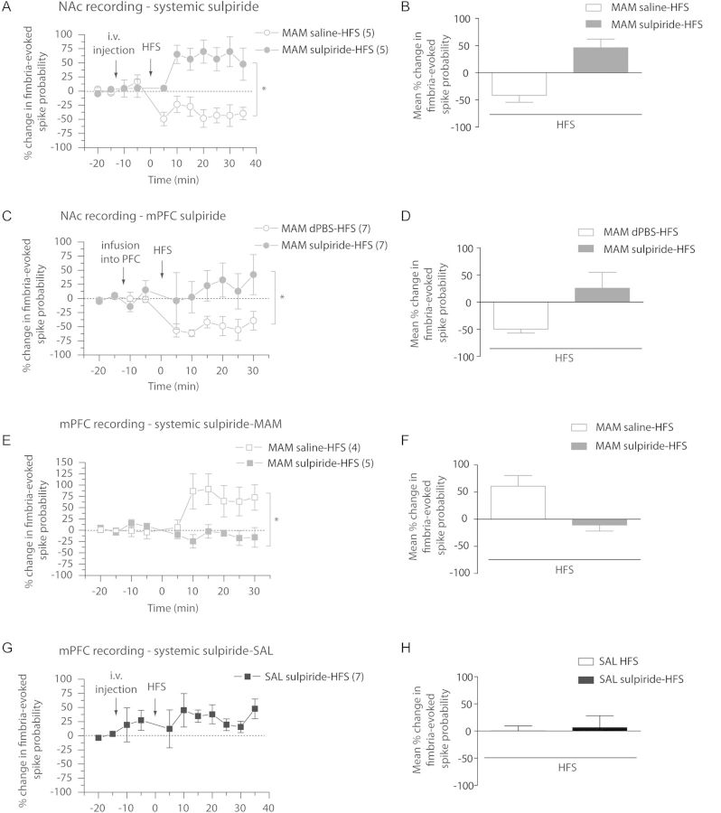
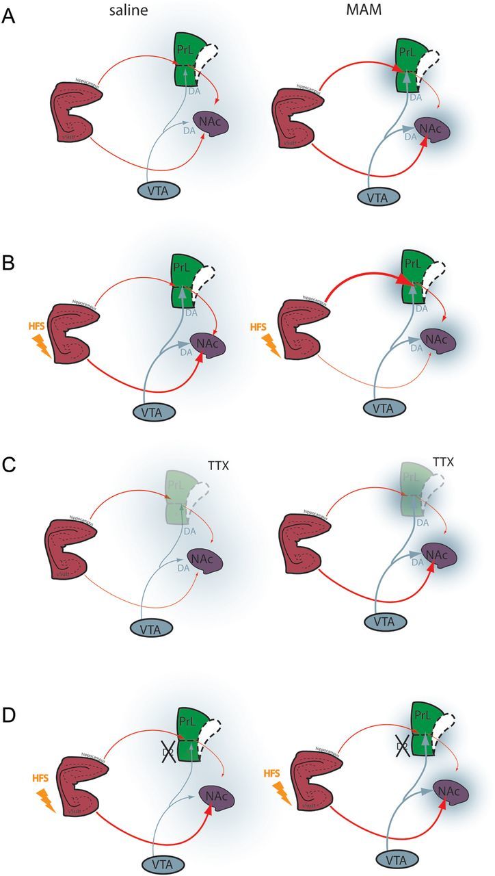
Schizophrenia is characterized by alterations in cortico-limbic processes believed to involve modifications in activity within the prefrontal cortex (PFC) and the hippocampus. The nucleus accumbens (NAc) integrates information from these 2 brain regions and is involved in cognitive and psychomotor functions that are disrupted in schizophrenia, indicating an important role for this structure in the pathophysiology of this disorder. In this study, we used in vivo electrophysiological recordings from the NAc and the PFC of adult rats and the MAM developmental disruption rodent model of schizophrenia to explore the influence of the medial PFC on the hippocampal-accumbens pathway. We found that, in MAM-treated rats, tetanization of hippocampal inputs to the NAc produce opposite synaptic plasticity compared with controls, which is a consequence of alterations in the hippocampal-mPFC pathway. Moreover, we show that administration of the D2-receptor-blocking antipsychotic drug sulpiride either systemically or directly into the mPFC reverses the alterations in the MAM rat. Therefore, specific disruptions in cortical and hippocampal inputs in the MAM-treated rat abnormally alter plasticity in subcortical structures. Moreover, our results suggest that, in the presence of antipsychotic drugs, the disrupted plasticities are normalized, supporting a role for this mechanism in antipsychotic drug action in schizophrenia.

Keywords: dopamine; long-term potentiation; nucleus accumbens; prefrontal cortex; schizophrenia.


Figures

Figure 1.
Figure 1.
HFS, but not LFS of the fimbria has opposite effect on accumbens plasticity in saline- and MAM-treated animals. (A and B) Representative example of extracellular recordings from NAc neuron activity evoked by fimbria stimulation before and after HFS to the fimbria in saline-treated animals (A) and MAM-treated animals (B). Twenty-five overlaid consecutive traces are shown with the numbers demonstrating the number of evoked spikes for 25 stimulations. Scale: 10 mV, 2 ms. (C) Mean percent change (±SEM) in fimbria-evoked spike probability, normalized to the baseline, after HFS to the fimbria in saline-treated (black circles) and MAM-treated (gray circles) animals (*P < 0.05; arrow indicates the time of stimulation). (D) Mean percent change in fimbria-evoked responses (±SEM) following HFS in saline-treated (black bar) and MAM-treated (gray bar) animals. (E) Mean percent change (±SEM) in fimbria-evoked spike probability, normalized to the baseline, after LFS to the fimbria in saline-treated (black circles) and MAM-treated (gray circles) animals (arrow indicates time of stimulation). (F) Mean percent change in fimbria-evoked responses (±SEM) following LFS in saline-treated (black bar) and MAM-treated (gray bar) animals.
Figure 2.
Figure 2.
HFS of fimbria induces abnormal plasticity in the mPFC in MAM-treated animals. (A) Schematic showing the placements of recording electrodes in the mPFC for saline-treated (black triangles) and MAM-treated (gray squares) rats, shown as coronal sections of the rat brain, taken from Paxinos and Watson (Paxinos and Watson 1996). (B) Representative example of extracellular mPFC recordings showing the response of pyramidal neurons to single fimbria stimulation (1 mA) in saline-treated animals (left) and MAM-treated animals (right). Scale: 10 mV, 10 ms (C) Scatterplot showing the spike probability over 5min in saline- and MAM-treated rats (black triangles and gray squares, respectively) evoked by 1 mA stimulation. (D) Mean percent change (±SEM) in fimbria-evoked spike probability, normalized to baseline, following HFS to the fimbria in saline-treated (black triangles) and MAM-treated (gray squares) animals (*P < 0.05; arrow indicates time of stimulation). (E) Mean of the percent change in fimbria-evoked responses (±SEM) following HFS in saline-treated (black bar) and MAM-treated (gray bar) animals.
Figure 3.
Figure 3.
Inactivation of the mPFC has opposite effect in the NAc of saline- and MAM-treated animals. (A and B) Representative example of extracellular recordings from accumbens neurons evoked by fimbria stimulation before and after infusion of tetrodotoxin (TTX) in the mPFC in saline-treated animals (A) and MAM-treated animals (B). Twenty-five overlaid consecutive traces are shown with the numbers demonstrating the number of evoked spikes for 25 stimulations. Scale: 10 mV, 5 ms. (C) Mean percent change (±SEM) of the fimbria-evoked spike probability, normalized to the baseline, after infusion of TTX (filled circle) or dPBS (open circles) into mPFC in saline-treated (black circles) and MAM-treated (gray circles) animals (*P < 0.05; arrow indicates the time of infusion). (D) Mean of the percent change in fimbria-evoked responses (±SEM) following infusion of TTX in saline-treated (black bar) and MAM-treated (gray bar) animals.
Figure 4.
Figure 4.
Abnormal accumbens plasticity following HFS to fimbria is reversed in MAM-treated animals by inactivating the mPFC. (A) Mean percent change (±SEM) in fimbria-evoked spike probability, normalized to baseline, following tetanization of the fimbria and infusion of TTX (closed circles) or vehicle, dPBS (open circles) in the mPFC in MAM-treated animals (*P < 0.05; arrows indicate time of stimulation and infusion). (B) Mean of the percent change in fimbria-evoked responses (±SEM) following infusion of vehicle, dPBS (white bar), or TTX (gray bar) in MAM-treated animals.
Figure 5.
Figure 5.
Administration of the D2 antagonist, sulpiride, reverses vSub–NAc LTD, and vSub–mPFC LTP in MAM-treated rats. (A) Mean percent change (±SEM) in fimbria-evoked spike probability in NAc neurons, normalized to baseline, following i.v. injection of sulpiride (closed circles) or saline (open circles) and tetanization of the fimbria in MAM-treated animals (*P < 0.05; arrows indicate time of injection and stimulation). (B) Mean of the percent change in fimbria-evoked responses (±SEM) after HFS to fimbria following an injection of sulpiride (gray bar) or saline (white bar) in MAM-treated animals. (C) Mean percent change (±SEM) in fimbria-evoked spike probability in NAc neurons, normalized to baseline, following local infusion of sulpiride (closed circles) or dPBS (open circles) in the mPFC and tetanization of the fimbria in MAM-treated animals (*P < 0.05; arrows indicate time of infusion and stimulation). (D) Mean of the percent change in fimbria-evoked responses (±SEM) after HFS to fimbria following local infusion of sulpiride (gray bar) or dPBS (white bar) in mPFC in MAM-treated animals. (E) Mean percent change (±SEM) in fimbria-evoked spike probability in mPFC neurons, normalized to baseline, following i.v. injection of sulpiride (closed squares) or saline (open squares) and HFS to the fimbria in MAM-treated animals (*P < 0.05; arrows indicate time of injection and stimulation). (F) Mean of the percent change in fimbria-evoked responses (±SEM) following HFS of the fimbria after injection of sulpiride (gray bar) or saline (white bar) in MAM-treated animals. (G) Mean percent change (±SEM) in fimbria-evoked spike probability in mPFC neurons, normalized to baseline, following i.v. injection of sulpiride and HFS to the fimbria in Saline-treated animals SAL; arrows indicate time of injection and stimulation). (H) Mean of the percent change in fimbria-evoked responses in saline animals (±SEM) following HFS of the fimbria alone (white bar) or after injection of sulpiride (black bar).
Figure 6.
Figure 6.
Schematic illustrating vSub–mPFC–NAc interactions in saline-and MAM-treated animals. (A) In saline rats, there is a balance between vSub and mPFC inputs to the NAC modulated by dopamine afferents from the VTA. In the MAM rats, there is a disruption of this balance due to hyperactivity in the vSub, leading to a hyperdopaminergic state and hypofunctional mPFC. (B) Tetanization of the fimbria increases the vSub–NAc pathway in saline animals (left) but induced a decrease in vSub–NAc drive and a hyperactive vSub–mPFC pathway in MAM rats (right). (C) Inactivation of the mPFC reduces the vSub drive of NAc neurons in saline rats (left) whereas in MAM rats, it increases the drive, demonstrating pathological phasic antagonistic action of the vSub and the mPFC on NAc neurons in our model. (D) Infusion of antipsychotic drug has no effect on the vSub–NAc HFS-induced synaptic plasticity in saline rats (Belujon and Grace 2008) (left) but normalizes the vSub–NAc and vSub–mPFC pathological synaptic plasticity in MAM rats (right). vSub, ventral subiculum of the hippocampus; PL, prelimbic area of the mPFC; IL, infralimbic area of the mPFC; NAc, nucleus accumbens; VTA, ventral tegmental area; DA, dopamine; TTX, tetrodotoxin; HFS, high-frequency stimulation.

References

    1. Akil M, Pierri JN, Whitehead RE, Edgar CL, Mohila C, Sampson AR, Lewis DA. Lamina-specific alterations in the dopamine innervation of the prefrontal cortex in schizophrenic subjects. Am J Psychiatry. 1999;156:1580–1589. - PubMed
    1. Belujon P, Grace AA. Critical role of the prefrontal cortex in the regulation of hippocampus-accumbens information flow. J Neurosci. 2008;28:9797–9805. - PMC - PubMed
    1. Benes FM, Berretta S. GABAergic interneurons: implications for understanding schizophrenia and bipolar disorder. Neuropsychopharmacology. 2001;25:1–27. - PubMed
    1. Flagstad P, Mork A, Glenthoj BY, van Beek J, Michael-Titus AT, Didriksen M. Disruption of neurogenesis on gestational day 17 in the rat causes behavioral changes relevant to positive and negative schizophrenia symptoms and alters amphetamine-induced dopamine release in nucleus accumbens. Neuropsychopharmacology. 2004;29:2052–2064. - PubMed
    1. Floresco SB, Blaha CD, Yang CR, Phillips AG. Modulation of hippocampal and amygdalar-evoked activity of nucleus accumbens neurons by dopamine: cellular mechanisms of input selection. J Neurosci. 2001;21:2851–2860. - PMC - PubMed

Publication types

MeSH terms