

Diagnosis and treatment of enthesitis-related arthritis
- PMID: 23236258
- PMCID: PMC3518441
- DOI: 10.2147/AHMT.S25872
Diagnosis and treatment of enthesitis-related arthritis
Abstract
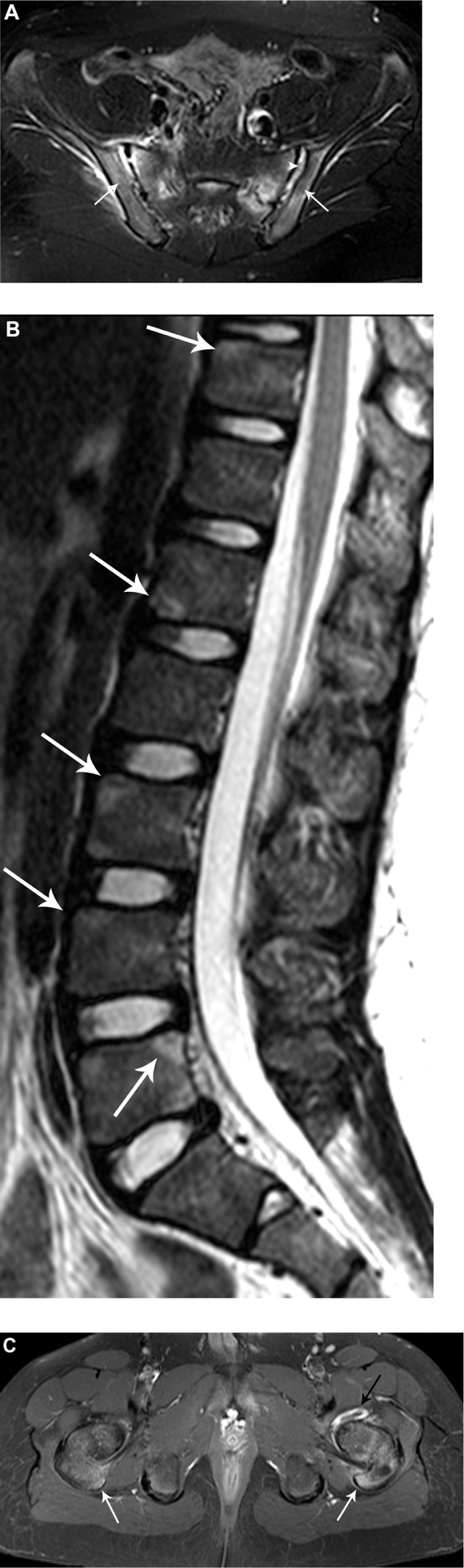
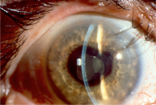
Juvenile idiopathic arthritis (JIA) is a chronic, inflammatory disease of unknown etiology. The enthesitis-related arthritis (ERA) JIA category describes a clinically heterogeneous group of children including some who have predominately enthesitis, enthesitis and arthritis, juvenile ankylosing spondylitis, or inflammatory bowel disease-associated arthropathy. ERA accounts for 10%-20% of JIA. Common clinical manifestations of ERA include arthritis, enthesitis, and acute anterior uveitis. Axial disease is also common in children with established ERA. Treatment regimens for ERA, many of them based on adults with rheumatoid arthritis and ankylosing spondylitis, include the use of nonsteroidal anti-inflammatory drugs, disease-modifying antirheumatic drugs, and biologic agents either individually or in combination.
Figures
References
-
- Karvonen M, Viik-Kajander M, Moltchanova E, Libman I, LaPorte R, Tuomilehto J. Incidence of childhood type 1 diabetes worldwide. Diabetes Mondiale (DiaMond) Project Group. Diabetes Care. 2000;23(10):1516–1526. - PubMed
-
- Solau-Gervais E, Robin C, Gambert C, et al. Prevalence and distribution of juvenile idiopathic arthritis in a region of Western France. Joint Bone Spine. 2010;77(1):47–49. - PubMed
-
- Oen K, Tucker L, Huber AM, et al. Predictors of early inactive disease in a juvenile idiopathic arthritis cohort: results of a Canadian multicenter, prospective inception cohort study. Arthritis Rheum. 2009;61(8):1077–1086. - PubMed
-
- Yilmaz M, Kendirli SG, Altintas DU, Karakoc GB, Inal A, Kilic M. Juvenile idiopathic arthritis profile in Turkish children. Pediatr Int. 2008;50(2):154–158. - PubMed
-
- Saurenmann RK, Rose JB, Tyrrell P, et al. Epidemiology of juvenile idiopathic arthritis in a multiethnic cohort: ethnicity as a risk factor. Arthritis Rheum. 2007;56(6):1974–1984. - PubMed
Grants and funding
LinkOut - more resources
Full Text Sources
Research Materials
