. 2012;7(12):e49708.
doi: 10.1371/journal.pone.0049708. Epub 2012 Dec 7.

Vasopressin inhibits LTP in the CA2 mouse hippocampal area

Affiliations

Vasopressin inhibits LTP in the CA2 mouse hippocampal area

Magda Chafai et al. PLoS One. 2012.

Abstract

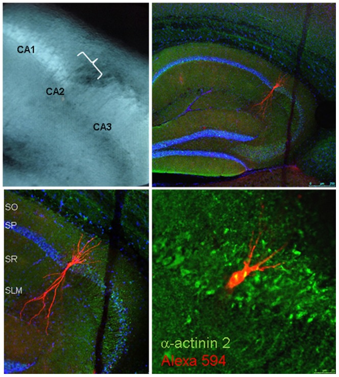
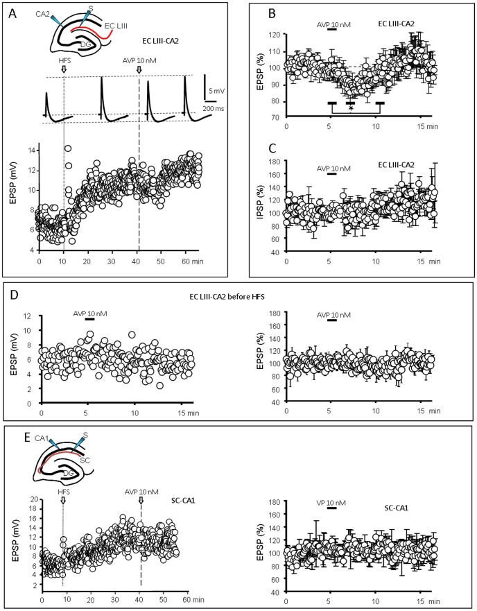
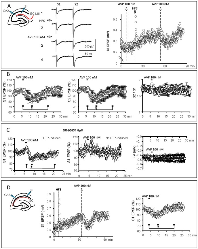
Growing evidence points to vasopressin (AVP) as a social behavior regulator modulating various memory processes and involved in pathologies such as mood disorders, anxiety and depression. Accordingly, AVP antagonists are actually envisaged as putative treatments. However, the underlying mechanisms are poorly characterized, in particular the influence of AVP on cellular or synaptic activities in limbic brain areas involved in social behavior. In the present study, we investigated AVP action on the synapse between the entorhinal cortex and CA2 hippocampal pyramidal neurons, by using both field potential and whole-cell recordings in mice brain acute slices. Short application (1 min) of AVP transiently reduced the synaptic response, only following induction of long-term potentiation (LTP) by high frequency stimulation (HFS) of afferent fibers. The basal synaptic response, measured in the absence of HFS, was not affected. The Schaffer collateral-CA1 synapse was not affected by AVP, even after LTP, while the Schaffer collateral-CA2 synapse was inhibited. Although investigated only recently, this CA2 hippocampal area appears to have a distinctive circuitry and a peculiar role in controlling episodic memory. Accordingly, AVP action on LTP-increased synaptic responses in this limbic structure may contribute to the role of this neuropeptide in controlling memory and social behavior.


Conflict of interest statement

Competing Interests: The authors have declared that no competing interests exist.

Figures

Figure 1
Figure 1. Recorded neurons are localized in the CA2 area and display a typical dendritic morphology.
Upper left: Bright field image of the hippocampal slice as seen during the experiment. Note that the CA2 is well delimited between the large pyramidal layer and mossy fiber tract of the CA3 area on the right and the thin pyramidal layer of the CA1 area on the left. Upper right: Fluorescence image (×5) of a neuron filled with Alexa-594-cadaverine (Red) during whole-cell recording and typically located in the CA2 area. Blue: Hoechst labeling of cell nuclei. Lower left: Higher magnification (×10) confirms that the injected neuron in located in the characteristic dilatation of the pyramidal layer in the CA2 area and displays the typical morphological characteristics of a pyramidal CA2 neuron, with few dendritic branches in stratum oriens (SO) and a dense branching in stratum lacunosum molecular (SLM). Lower right: Merged high magnification (×40) fluorescence image showing in red the Alexa-594-cadaverine filling the injected neuron and in green the immunolabeling of α-actinin2, a protein enriched in CA2 neurons. Note the co-localization of cadaverin and α-actinin, demonstrating that the recorded neuron was located in the CA2 area. SO, stratum oriens; SP, stratum pyramidale; SR, stratum radiatum; SLM, stratum lacunosum moleculare.
Figure 2
Figure 2. Vasopressin decreases EPSPs in CA2 after LTP induction.
A: Top: typical recordings of the combined EPSP/IPSP evoked by stimulation of LIII entorhinal fibers (EC LIII) and recorded in CA2 pyramidal neurons (inset). Traces are averages of 12 successive responses recorded during 1 min, before high frequency stimulation (HFS) and during LTP before, during and after the response to AVP (10 nM ; 1 min). Bottom graph: typical example showing that the amplitude of the EPSP increased transiently (short-term facilitation) after HFS and then progressively to reach a plateau (long-term potentiation: LTP). Vasopressin (AVP 10 nM; 1 min) applied during LTP transiently decreased the EPSP amplitude. B) Average graph of EPSP amplitude vs. time (n = 6; * p<0.05) demonstrating that AVP inhibited the CA2 EPSP evoked by EC LIII stimulation during LTP. Values are expressed as % of the mean of the responses recorded during 5 min before AVP application. C) Average graph (n = 6) showing that the IPSP component of the CA2 response to EC LIII stimulation was not affected by AVP. D) Typical graph (Left) and average graph (Right, n = 10) of EPSP amplitude vs. time showing that AVP did not affect the basal EC LIII-CA2 EPSP recorded before HFS stimulation. E) Left, Typical graph of the fEPSP evoked by Schaffer collaterals stimulation and recorded in a CA1 pyramidal neuron (SC-CA1); AVP did not affect the amplitude of the LTP-potentiated EPSP , as shown on the average graph (Right, n = 4).
Figure 3
Figure 3. Vasopressin decreased the population fEPSP evoked by stimulation of LIII entorhinal fibers (EC LIII) and recorded in the CA2 dendritic field.
A) Left : Typical recordings (left, 12 traces averaged over 1 min) of fEPSPs evoked by two successive stimulations before HFS (1) and during LTP before (2), during (3) and after (4) the response to AVP (100 nM ; 1 min). Note that the response to the second stimulation (S2) is larger than the first one (S1), indicative of a paired-pulse facilitation. Right, corresponding graph of the fEPSP amplitude vs. time. HFS induced a short- and then a long-term potentiation. Values correspond to S1. AVP (1 min) transiently decreased the LTP-potentiated fEPSP. B) Average graphs (n = 8; values expressed as % of the mean of the responses recorded during 5 min before AVP application) demonstrating that AVP (100 nM; 1 min) transiently and significantly decreased the amplitude of both S1 and S2 fEPSPs without affecting the paired-pulse facilitation (calculated as the ratio S2/S1). C) Left: fEPSP recorded in the presence of the GABAA antagonist SR 95531. AVP was effective to decrease the fEPSP during LTP (n = 14) but failed to affect the fEPSP in slices in which no LTP was evoked (Middle; n = 7). Right, Absence of effect of AVP on the fiber volley (FV) amplitude (n = 5), demonstrating that AVP did not reduce the excitability of the EC LIII fibers. D) fEPSP recorded in the CA2 dendritic area during stimulation of SC fibers. Left, Typical recording in a slice in which HFS induced a LTP and AVP reduced the potentiated fEPSP. Right, Average graph (n = 7) demonstrating that AVP (100 nM; 1 min) transiently and significantly decreased the amplitude of the SC-CA2 fEPSPs.

References

    1. Van Wimersma Greidanus TB, De Wied D (1980) Physiological significance of neurohypophyseal hormones in memory processes. Acta Psychiatr Belg 80: 721–727. - PubMed
    1. Bao AM, Meynen G, Swaab DF (2008) The stress system in depression and neurodegeneration: focus on the human hypothalamus. Brain Res Rev 57: 531–553. - PubMed
    1. Frank E, Landgraf R (2008) The vasopressin system–from antidiuresis to psychopathology. Eur J Pharmacol 583: 226–242. - PubMed
    1. Surget A, Belzung C (2008) Involvement of vasopressin in affective disorders. Eur J Pharmacol 583: 340–349. - PubMed
    1. Caldwell HK, Lee HJ, Macbeth AH, Young WS 3rd (2008) Vasopressin: behavioral roles of an “original” neuropeptide. Prog Neurobiol 84: 1–24. - PMC - PubMed

Publication types

MeSH terms