. 2012;7(12):e51259.
doi: 10.1371/journal.pone.0051259. Epub 2012 Dec 7.

GTSE1 is a microtubule plus-end tracking protein that regulates EB1-dependent cell migration

Affiliations

GTSE1 is a microtubule plus-end tracking protein that regulates EB1-dependent cell migration

Massimilano Scolz et al. PLoS One. 2012.

Abstract

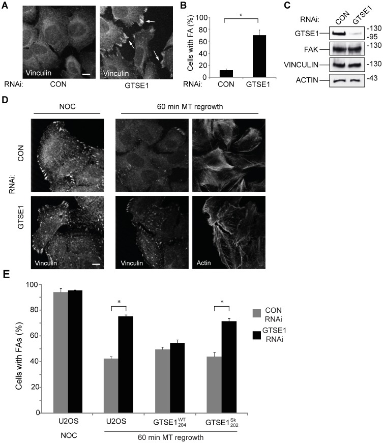
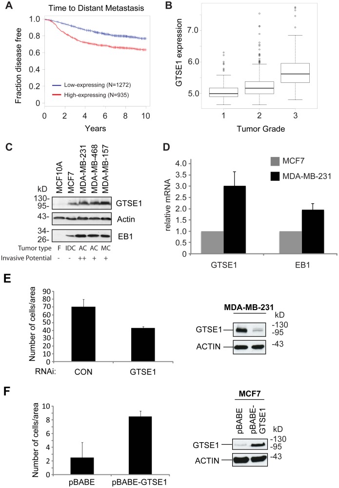
The regulation of cell migration is a highly complex process that is often compromised when cancer cells become metastatic. The microtubule cytoskeleton is necessary for cell migration, but how microtubules and microtubule-associated proteins regulate multiple pathways promoting cell migration remains unclear. Microtubule plus-end binding proteins (+TIPs) are emerging as important players in many cellular functions, including cell migration. Here we identify a +TIP, GTSE1, that promotes cell migration. GTSE1 accumulates at growing microtubule plus ends through interaction with the EB1+TIP. The EB1-dependent +TIP activity of GTSE1 is required for cell migration, as well as for microtubule-dependent disassembly of focal adhesions. GTSE1 protein levels determine the migratory capacity of both nontransformed and breast cancer cell lines. In breast cancers, increased GTSE1 expression correlates with invasive potential, tumor stage, and time to distant metastasis, suggesting that misregulation of GTSE1 expression could be associated with increased invasive potential.


Conflict of interest statement

Competing Interests: The authors have declared that no competing interests exist.

Figures

Figure 1
Figure 1. GTSE1 is an EB1-dependent microtubule plus end growth tracking protein.
(A) Still images of a live U2OS cell stably expressing GTSE1-GFP and mCherry-alpha-tubulin from Movie S1. GTSE1-GFP is enriched at growing microtubule plus ends, and also associated with the microtubule lattice. (B) Western blot showing GTSE1 and EB1 levels after EB1 RNAi. U2OS cells stably expressing GFP-GTSE1 were transfected with a control (siCONT) or EB1 (siEB1) siRNA for 36 h. Blots were probed with antibodies against GFP, EB1 or actin (loading control). (C) Still images of live U2OS cells expressing GTSE1-GFP after transfection with control (CON) or EB1 siRNA, from Movies S2 and S3. After EB1 depletion, GTSE1-GFP no longer tracks growing microtubule ends, but remains associated with the microtubule lattice.
Figure 2
Figure 2. GTSE1 is recruited to microtubule plus ends through short EB1-interaction motifs.
(A) Sequence alignment of hGTSE1 amino acids 503–538 that contain tandem conserved SKIP-like motifs. The first four rows contain GTSE1 homologs from human (hs), mouse (mm), Xenopus (xl) and zebrafish (dr). The last three rows show conserved regions from other human +TIPs. SKIP-like motifs are highlighted in green boxes. Conserved TP motifs are highlighted in grey boxes. Basic residues are colored red, serines and threonines are colored blue. (B) GTSE1 immunoprecipitates EB1 in U2OS cells. U2OS cell lysates were immunoprecipitated with anti-GTSE1 antibody, or anti-GFP as a control. Input lysate and immunoprecipitated fractions were run by SDS-PAGE and Western blotted with either anti-GTSE1 or anti-EB1 antibody. (C) In vitro pull-down binding assay using purified GST or GST-EB1 fusion proteins incubated with in vitro translated 35S-labeled GTSE1 WT (hGTSE1 WT IVT) or GTSE1 mutated at the SKIP motifs (L511N P512N L522N P523N) (GTSE1 Sk IVT). Inputs represent 20% of IVTs used for pull-down assays. The top gel shows IVT GTSE1 by autoradiograph, bottom gels are commassie stained. GST-EB1 interacts with in vitro translated GTSE1, but not GTSE1 mutated at SKIP motifs. (D) Still images of live clonal U2OS cells expressing wild type GTSE1-GFP (GTSE1WT 204) or GTSE1-GFP mutated at the SKIP motifs (L511N P512N L522N P523N) (GTSE1Sk 202)(Movie S4). Similar to EB1 depletion, the mutated GTSE1-GFP does not track growing microtubule tips, but localizes to the microtubule lattice. All scale bars represent 10 microns.
Figure 3
Figure 3. GTSE1 binds directly to the microtubule lattice and stops tip tracking in mitosis.
(A) Microtubule co-sedimentation assay. In vitro translated 35S-labeled GTSE1 (GTSE1 IVT) was incubated with taxol-stabilized microtubules (MTs +) or with taxol-containing buffer (without microtubules; MTs −). Supernatant (SN) and pellet (P) fractions were separated by SDS-PAGE and the presence of GTSE1 in each fraction was detected by autoradiography. (B) Still images of a TIRF field showing GTSE1-GFP (left panel) binding to rhodamine-labeled GMPCPP seeds (right panel) and along the growing MT lattice. Scale bar represents 2 microns. (C) Still images of live U2OS cells stably expressing GTSE1-GFP and EB3-mCherry with DNA labeled with Hoechst, from Movie S5. Time GTSE1-GFP tracks growing plus ends in prophase, but not 8 minutes later in prometaphase. Scale bar represents 10 microns. (D) Western blot of endogenous GTSE1 in Xenopus meiotic (M) and interphase (I) extract. (E) Still images of TIRF fields and representative kymographs of GMPCPP-stabilized microtubules incubated with interphase (Movie S7) or mitotic (Movie S8) Xenopus egg extracts and purified hGTSE1-GFP protein. Horizontal scale bar represents 5 microns, vertical scale bar 10 seconds. (F) Still images of TIRF fields and representative kymographs of GMPCPP-stabilized microtubules incubated with mitotic Xenopus egg extracts and purified hGTSE1-GFP or EB1-GFP protein, after addition of purified cyclinBΔ90, from Movies S9 and S10.
Figure 4
Figure 4. GTSE1 modulates cell migration in an EB1-dependent manner.
(A) Transwell migration assay and western blot in U2OS cells, and U2OS cells stably expressing RNAi-resistant wild-type GTSE1-GFP (GTSE1WT 204), or RNAi-resistant GTSE1-GFP mutated at the SKIP motifs (GTSE1Sk 202). Cells were transfected with control (CON) or GTSE1 siRNA for 36 h and seeded on transwell membranes. Histograms show the mean number of cells/area that migrated through the transwell after 16 h (10 replicates/experiment). Error bars represent the standard error of the mean from three independent experiments. Western blots were performed on cells after the same treatment, and blotted with anti-GTSE1 and anti-actin. (B) Transwell migration assay and western blot in Wi38 cells. Cells were transfected and analysed as in (a). (C) Transwell migration assay and western blot in HCT116 wild type, HCT116 p53−/−, and HCT116 p21−/− cell lines. Cells were transfected and analyzed as in (a). Western blots were performed on cells after the same treatment, and blotted with anti-GTSE1, anti-p21, anti-p53, and anti-actin. (D) Transwell migration assay and western blot in H1299 cells containing inducible constructs for expression of wild type GTSE1 (GTSE1 WT) or SKIP-domain mutated GTSE1 (GTSE1 Sk). Cells were untreated (−) or treated with Ponasterone A (PonA) (+) for 24 h to induce GTSE1 expression, then trypsinized and seeded on transwell membranes. Histograms show the mean number of cells/area that migrated through the transwell after 16 h (10 replicates/experiment). Western blots were performed on cells after the same treatment, and blotted with anti-GTSE1 and anti-actin.
Figure 5
Figure 5. GTSE1 modulates focal adhesion disassembly in an EB1-dependent manner.
(A) Immunofluorescence of U2OS cells transfected with control (CON) or GTSE1 siRNA for 24 h followed by serum starvation for 48 h, stained for vinculin. Focal adhesions persist in cells depleted of GTSE1. Scale bar represents 10 microns. (B) Quantification of focal adhesion (FA) disassembly from experiments from (A). The percentage of cells containing 10 or more focal adhesions after serum starvation-induced disassembly was determined (n = >50 cells per experiment, 3 experiments for each condition). * indicates p<0.05 as determined by a Student’s t test. (C) Western blot of U2OS cells transfected with control (CON) or GTSE1 siRNA and serum-starved for 48 h, blotted with anti-GTSE1, anti-FAK, anti-vinculin, and anti-actin. (D) Immunofluorescence of U2OS cells transfected with control (CON) or GTSE1 siRNA for 36 hours. Cells were imaged after treatment with nocodazole for 4 hours, and 60 minutes following washout of nocodazole to allow microtubule regrowth. Cells are stained for vinculin and actin. GTSE1-depleted cells contain more focal adhesions that wild-type following microtubule regrowth. Scale bar represents 10 microns. (E) Quantification of focal adhesion disassembly in U2OS cells, following the assay described in (D). Cells stably expressing RNAi-resistant wild-type GTSE1-GFP (GTSE1WT 204) or GTSE1-GFP mutated at the SKIP motifs (GTSE1Sk 202) were additionally assayed. Quantification was performed as described in (B). Cells containing only mutant GTSE1 unable to interact with EB1 or track growing microtubule ends are deficient for microtubule-dependent focal adhesion disassembly.
Figure 6
Figure 6. GTSE1 expression in breast cancer tumors and cells correlates with time to metastasis and invasiveness.
(A) Kaplan–Meier survival curve of time to distant metastasis of breast cancer patients classified according to the expression of GTSE1. Red line: cases with high expression of GTSE1, blue line: cases with low expression of GTSE1. (p-value <10?–15) (B) Boxplots of the distribution of gene expression intensities of GTSE1 across different breast cancer subtypes (Grade 1, 2 or 3; p<10?-5; linear regression analysis),. (C) Western blot analysis of GTSE1 and EB1 protein levels in different breast cancer cell lines. Tumor types are: F, fibrocystic disease, non-transformed, immortal cell line; IDC, invasive ductal carcinoma; AC, adenocarcinoma; MC, metaplastic carcinoma. Invasive potential is characterized as not invasive (−), invasive (+), or highly invasive (++). Adapted from Neve et al. Cancer Cell 2006. (D) Quantitative RT-PCR analysis of GTSE1 and EB1 relative mRNA levels in MCF7 and MDA-MB-231 cells. Error bars represent the standard error of the mean from three independent experiments. p<0.01 (Student’s t-test). (E) Transwell migration assay and western blot of the MDA-MB-231 cell line. Cells were transfected with control (CON) or GTSE1 siRNA for 36 hours, trypsinized, and seeded on transwell membranes. Histograms show the mean number of cells/area that migrated through the transwell after 16 h (10 replicates/experiment). Error bars represent the standard error of the mean from three independent experiments. * indicates p<0.05 (Student’s t-test). Western blots were performed on cells after the same treatment, and blotted with anti-GTSE1 and anti-actin. (F) Transwell migration assay and western blot of the MCF7 cell line containing a stably integrated GTSE1 overexpression construct (pBABE-GTSE1) or empty vector (pBABE). Cells were trypsinized and seeded on transwell membranes. Histograms show the mean number of cells/area that migrated through the transwell after 16 h (10 replicates/experiment). Western blots were performed on cells after the same treatment, and blotted with anti-GTSE1 and anti-actin.

References

    1. Zhao X, Guan J-L (2011) Focal adhesion kinase and its signaling pathways in cell migration and angiogenesis. Advanced Drug Delivery Reviews 63: 610–615 doi:10.1016/j.addr.2010.11.001. - DOI - PMC - PubMed
    1. Li D-M, Feng Y-M (2011) Signaling mechanism of cell adhesion molecules in breast cancer metastasis: potential therapeutic targets. Breast Cancer Res Treat 128: 7–21 doi:10.1007/s10549-011-1499-x. - DOI - PubMed
    1. Thiery JP (2002) Epithelial–mesenchymal transitions in tumour progression. Nat Rev Cancer 2: 442–454 doi:10.1038/nrc822. - DOI - PubMed
    1. Palmer TD, Ashby WJ, Lewis JD, Zijlstra A (2011) Targeting tumor cell motility to prevent metastasis. Advanced Drug Delivery Reviews 63: 568–581 doi:10.1016/j.addr.2011.04.008. - DOI - PMC - PubMed
    1. Kaverina I, Straube A (2011) Regulation of cell migration by dynamic microtubules. Seminars in Cell and Developmental Biology 22: 968–974 doi:10.1016/j.semcdb.2011.09.017. - DOI - PMC - PubMed

Publication types

MeSH terms