Comparative Study
. 2012;7(12):e51310.
doi: 10.1371/journal.pone.0051310. Epub 2012 Dec 7.

Localized intestinal radiation and liquid diet enhance survival and permit evaluation of long-term intestinal responses to high dose radiation in mice

Affiliations
Comparative Study

Localized intestinal radiation and liquid diet enhance survival and permit evaluation of long-term intestinal responses to high dose radiation in mice

Laurianne Van Landeghem et al. PLoS One. 2012.

Abstract

Background: In vivo studies of high dose radiation-induced crypt and intestinal stem cell (ISC) loss and subsequent regeneration are typically restricted to 5-8 days after radiation due to high mortality and immune failure. This study aimed to develop murine radiation models of complete crypt loss that permit longer-term studies of ISC and crypt regeneration, repair and normalization of the intestinal epithelium.

Methods: In C57Bl/6J mice, a predetermined small intestinal segment was exteriorized and exposed to 14 Gy-radiation, while a lead shield protected the rest of the body from radiation. Sham controls had segment exteriorization but no radiation. Results were compared to C57Bl/6J mice given 14 Gy-abdominal radiation. Effects of elemental liquid diet feeding from the day prior to radiation until day 7 post-radiation were assessed in both models. Body weight and a custom-developed health score was assessed every day until day 21 post-radiation. Intestine was assessed histologically.

Results: At day 3 after segment radiation, complete loss of crypts occurred in the targeted segment, while adjacent and remaining intestine in segment-radiated mice, and entire intestine of sham controls, showed no detectable epithelial damage. Liquid diet feeding was required for survival of mice after segment radiation. Liquid diet significantly improved survival, body weight recovery and normalization of intestinal epithelium after abdominal radiation. Mice given segment radiation combined with liquid diet feeding showed minimal body weight loss, increased food intake and enhanced health score.

Conclusions: The segment radiation method provides a useful model to study ISC/crypt loss and long-term crypt regeneration and epithelial repair, and may be valuable for future application to ISC transplantation or to genetic mutants that would not otherwise survive radiation doses that lead to complete crypt loss. Liquid diet is a simple intervention that improves survival and facilitates long-term studies of intestine in mice after high dose abdominal or segment radiation.


Conflict of interest statement

Competing Interests: The authors have declared that no competing interests exist.

Figures

Figure 1
Figure 1. The segment radiation method allows specific and total crypt ablation in a pre-determined intestinal segment.
A. Illustrative photograph of H&E-stained Swiss-rolled jejunum from sham controls shows that no damage was induced by the segment exteriorization procedure (n = 3). Scale Bar: 500 µm. B. The schematic represents Swiss-rolled jejunum (black spiral). The radiated segment is shown in red between the arrow head and the arrow. The 3 rectangles (C, D and E) illustrate the localization of the radiated segment in the photographs shown in C, D and E. Illustrative photographs of H&E-stained Swiss-rolled jejunum collected from a mouse at day 3 post-radiation demonstrates that the segment radiation procedure induced specific and complete ablation of crypts in the targeted portion of the jejunum delimited by the arrowhead and the arrow, while the rest of the tissue showed normal epithelial architecture (n = 3). In these experiments, all mice (segment-radiated or sham controls) received liquid diet.
Figure 2
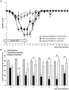
Figure 2. Liquid diet leads to maximal survival after both segment and abdominal radiation.
Graphs show percent survival at different times after abdominal or segment radiation in mice given normal chow or liquid diet (from the day prior to radiation until day 7 post-radiation). 52.9% of the mice fed with normal chow and abdominally irradiated survived for 21 days post-radiation while none survived when fed with normal chow and locally radiated on a determined intestinal segment. Liquid diet treatment significantly improved survival after both local and abdominal radiation (n≥6: Abdominal Radiation-Normal Chow n = 17; Abdominal Radiation-Liquid Diet n = 14; Segment Radiation-Normal Chow n = 6; Segment Radiation-Liquid Diet n = 14; Mann-Whitney; a: p<0.05 vs. Abdominal Radiation-Normal Chow; b: p<0.05 vs. Abdominal Radiation-Liquid Diet; c: p<0.05 vs. Segment Radiation-Liquid Diet).
Figure 3
Figure 3. The segment radiation method combined with liquid diet induces minimal body weight loss.
Graphs show percent body weight loss (mean ± SEM) at different times after irradiation in abdominally-radiated mice given normal chow or liquid diet and in segment-radiated mice given liquid diet. Mice locally radiated on an intestinal segment and fed with liquid diet exhibited minimal body weight loss and rapid body weight recovery after radiation compared with mice abdominally radiated. Liquid diet significantly improved body weight recovery of mice treated with abdominal radiation (n≥9: Abdominal Radiation-Normal Chow n = 12; Abdominal Radiation-Liquid Diet n = 9; Segment Radiation-Liquid Diet n = 9; Two Way ANOVA; a: p<0.05 vs. Abdominal Radiation-Liquid Diet; b: p<0.05 vs. Abdominal Radiation-Normal Chow).
Figure 4
Figure 4. The segment radiation method combined with liquid diet improves health post-radiation.
A. Graphs show health scores (mean ± SEM) of abdominally-radiated mice given normal chow or liquid diet and segment-radiated mice given liquid diet at different times after irradiation. Mice locally radiated on a targeted intestinal segment and fed with liquid diet exhibited significantly improved health score between day 4 and day 8 post-radiation as compared with abdominally radiated mice. Liquid diet tends to slightly improve health score between day 4 and day 8 post abdominal radiation (n≥8: Abdominal Radiation-Normal Chow n = 11; Abdominal Radiation-Liquid Diet n = 8; Segment Radiation-Liquid Diet n = 8; ANOVA; a: p<0.05 vs. Abdominal Radiation-Liquid Diet; b: p<0.05 vs. Abdominal Radiation-Normal Chow). B. Bar Graphs show amounts of liquid diet consumed per mouse per day in ml (mean ± SEM) in non-irradiated mice or mice given abdominal or segment radiation. Mice locally radiated on an intestinal segment exhibited significantly increased intake of liquid diet as compared with abdominally-radiated mice and had similar intake to non-irradiated control mice by 3–5 days after irradiation (n≥12: No Radiation n = 13; Abdominal Radiation n = 14; Segment Radiation n = 12; Mann-Whitney; a: p<0.05 vs. No Radiation; b: p<0.05 vs. Abdominal Radiation; NS: not significant).
Figure 5
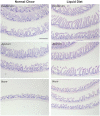
Figure 5. Liquid diet promotes normalization of the intestinal epithelium after abdominal radiation.
H&E staining of Swiss-rolled intestines at day 21 post-abdominal radiation showed enlarged crypts as well as blunted villi in mice fed with normal chow and overall more abnormal mucosal epithelium when compared with mice fed with liquid diet (n = 3). Scale Bar: 500 µm.
Figure 6
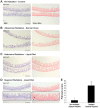
Figure 6. Locally-radiated intestinal segments exhibit mild fibrosis at day 21 post-radiation.
A–C. H&E staining (left panels) and Sirius red staining (right panels) revealed no evident sign of fibrosis in distal jejunum of abdominally-radiated mice fed with normal chow (B) or liquid diet (C) at day 21 post-radiation as compared to non-irradiated controls (A). Scale Bar: 500 µm. D. Left panel. However, in mice given segment radiation, the radiated segment (black rectangle) was still distinguishable from adjacent non-irradiated intestine at day 21 post-radiation by H&E staining and exhibited a thickening of the muscular wall (double headed arrow). Right panel. Sirius red staining revealed an increase in collagen deposition within the submucosa (arrow) and the serosa (arrowhead) of the radiated segment (black rectangle). Scale Bar: 500 µm. E. Quantification of the extent of the collagen deposition demonstrated a significant increase in fibrosis in the radiated segment vs. non-irradiated adjacent tissue at 21 days after irradiation (n = 3; paired t-test; a: p<0.05).

Similar articles

Cited by

References

    1. Williams JP, Brown SL, Georges GE, Hauer-Jensen M, Hill RP, et al. (2010) Animal models for medical countermeasures to radiation exposure. Radiat Res 173: 557–578. - PMC - PubMed
    1. Somosy Z, Horvath G, Telbisz A, Rez G, Palfia Z (2002) Morphological aspects of ionizing radiation response of small intestine. Micron 33: 167–178. - PubMed
    1. Theis VS, Sripadam R, Ramani V, Lal S (2010) Chronic radiation enteritis. Clin Oncol (R Coll Radiol) 22: 70–83. - PubMed
    1. Zimmerer T, Bocker U, Wenz F, Singer MV (2008) Medical prevention and treatment of acute and chronic radiation induced enteritis–is there any proven therapy? a short review. Z Gastroenterol 46: 441–448. - PubMed
    1. van der Flier LG, Clevers H (2009) Stem cells, self-renewal, and differentiation in the intestinal epithelium. Annu Rev Physiol 71: 241–260. - PubMed

Publication types