. 2012;7(12):e51331.
doi: 10.1371/journal.pone.0051331. Epub 2012 Dec 7.

The anti-proliferative activity of BTG/TOB proteins is mediated via the Caf1a (CNOT7) and Caf1b (CNOT8) deadenylase subunits of the Ccr4-not complex

Affiliations

The anti-proliferative activity of BTG/TOB proteins is mediated via the Caf1a (CNOT7) and Caf1b (CNOT8) deadenylase subunits of the Ccr4-not complex

Rachel Doidge et al. PLoS One. 2012.

Abstract

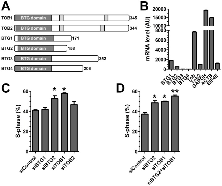
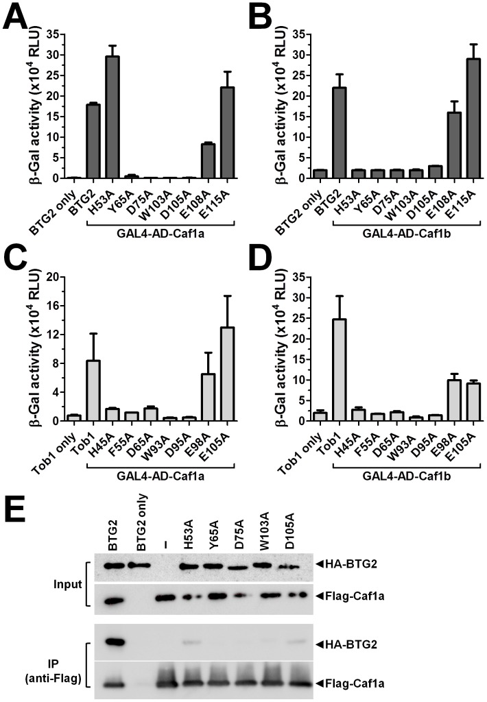
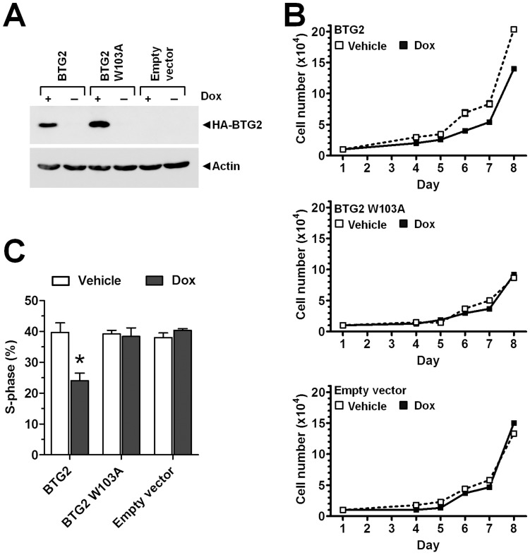
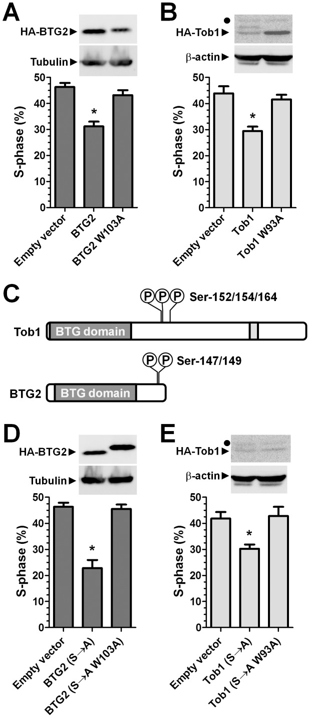
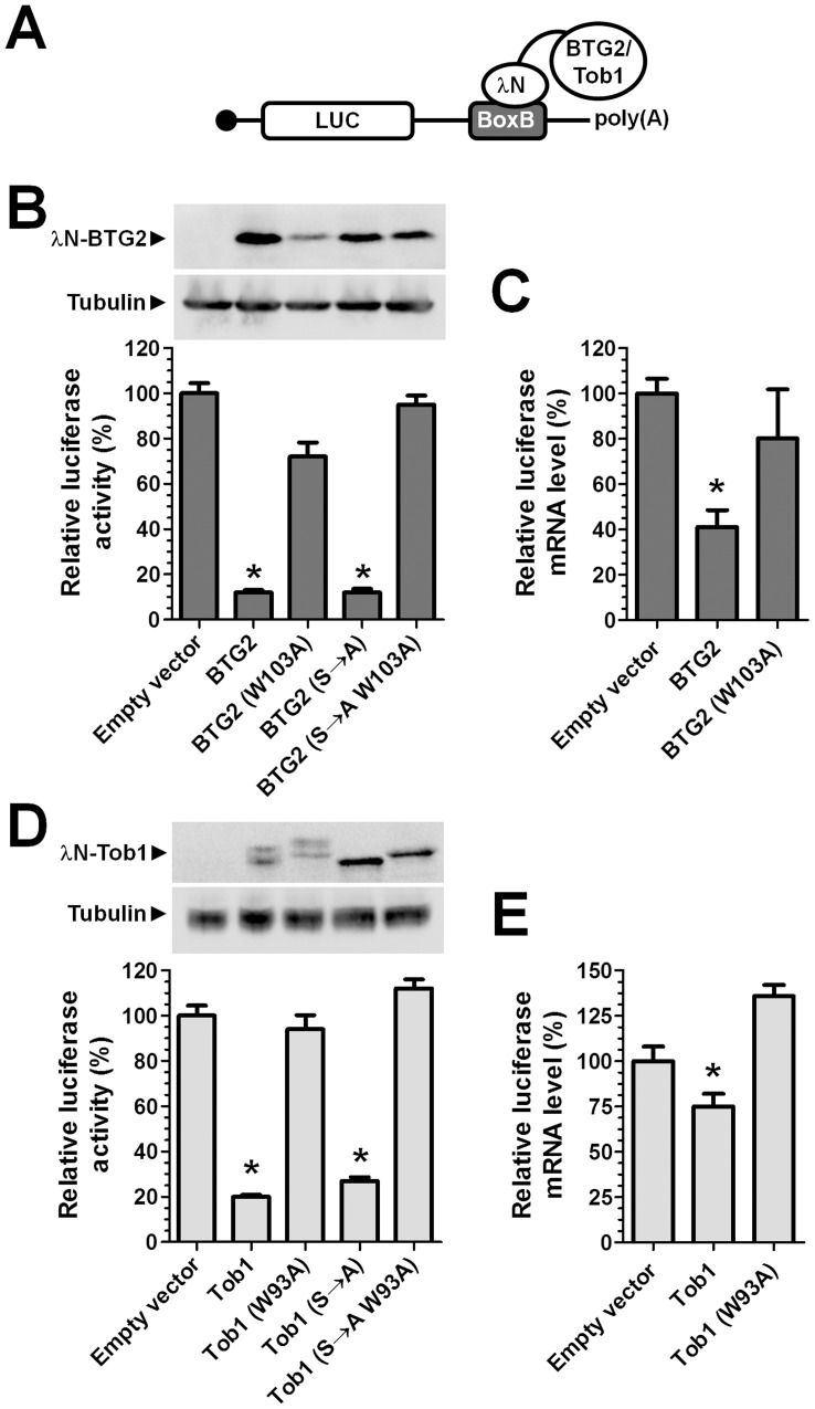
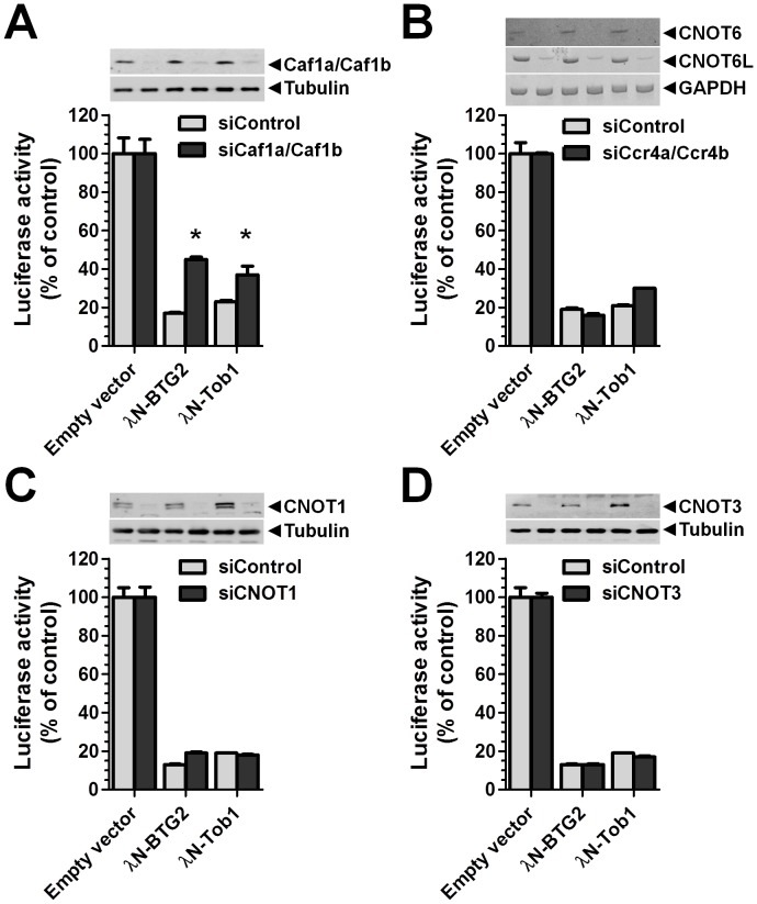
The human BTG/TOB protein family comprises six members (BTG1, BTG2/PC3/Tis21, BTG3/Ana, BTG4/PC3B, TOB1/Tob, and TOB2) that are characterised by a conserved BTG domain. This domain mediates interactions with the highly similar Caf1a (CNOT7) and Caf1b (CNOT8) catalytic subunits of the Ccr4-Not deadenylase complex. BTG/TOB proteins have anti-proliferative activity: knockdown of BTG/TOB can result in increased cell proliferation, whereas over-expression of BTG/TOB leads to inhibition of cell cycle progression. It was unclear whether the interaction between BTG/TOB proteins and the Caf1a/Caf1b deadenylases is necessary for the anti-proliferative activity of BTG/TOB. To address this question, we further characterised surface-exposed amino acid residues of BTG2 and TOB1 that mediate the interaction with the Caf1a and Caf1b deadenylase enzymes. We then analysed the role of BTG2 and TOB1 in the regulation of cell proliferation, translation and mRNA abundance using a mutant that is no longer able to interact with the Caf1a/Caf1b deadenylases. We conclude that the anti-proliferative activity of BTG/TOB proteins is mediated through interactions with the Caf1a and Caf1b deadenylase enzymes. Furthermore, we show that the activity of BTG/TOB proteins in the regulation of mRNA abundance and translation is dependent on Caf1a/Caf1b, and does not appear to require other Ccr4-Not components, including the Ccr4a (CNOT6)/Ccr4b (CNOT6L) deadenylases, or the non-catalytic subunits CNOT1 or CNOT3.


Conflict of interest statement

Competing Interests: The authors have declared that no competing interests exist.

Figures

Figure 1
Figure 1. Knockdown of BTG2 and TOB1 results in increased proliferation of MCF-7 cells.
(A) Overview of the human BTG/TOB family. The BTG domain is highlighted in grey, and PAM2 motifs present in TOB1 and TOB2 are highlighted in light grey. (B) Expression of BTG/TOB family members in MCF-7 cells. Data was obtained by genome-wide expression data using microarrays and confirmed by RT-qPCR. (C) Knockdown of BTG2 and TOB1 results in increased proliferation of MCF7 cells. Cells in S-phase were detected by labelling using the nucleoside analogue EdU 48 h after siRNA transfection. No effect was observed upon knockdown of BTG1 and TOB2. * p<0.05 (compared to treatment with non-targeting control siRNA). (D) Combined knockdown of BTG2 and TOB1 results in further increased cell proliferation of MCF7 cells. Cells in S-phase were detected by labelling using the nucleoside analogue EdU 48 h after siRNA transfection. * p<0.05 (compared to non-targeting control siRNA). ** p<0.05 (compared to BTG2 or TOB1 knockdown). All experiments were carried out in triplicate. Error bars represent the standard error of the mean.
Figure 2
Figure 2. Amino acid residues of BTG2 and TOB1 mediating interactions with the Caf1a and Caf1b deadenylase enzymes.
(A) Two-hybrid interaction analysis of BTG2 containing amino acid substitutions fused to the GAL4 DNA-binding domain and Caf1a fused to the activation domain of GAL4. (B) Two-hybrid interaction analysis of BTG2 containing amino acid substitutions fused to the GAL4 DNA-binding domain and Caf1b fused to the activation domain of GAL4. Plasmids encoding Gal4-DNA binding domain-BTG2 and Gal4-activation domain-Caf1a/Caf1b were transformed into yeast strain YRG-2 containing a LacZ reporter gene under control of five GAL4 consensus sequences. All experiments were carried out in triplicate. Error bars represent the standard error of the mean. (C) Two-hybrid interaction analysis of TOB1 containing amino acid substitutions fused to the GAL4 DNA-binding domain and Caf1a fused to the activation domain of GAL4. (D) Two-hybrid interaction analysis of TOB1 containing amino acid substitutions fused to the GAL4 DNA-binding domain and Caf1b fused to the activation domain of GAL4. Plasmids encoding Gal4-DNA binding domain-TOB1 and Gal4-activation domain-Caf1a/Caf1b were transformed into yeast strain YRG-2 containing a LacZ reporter gene under control of five GAL4 consensus sequences. All experiments were carried out in triplicate. Error bars represent the standard error of the mean. (E) Interaction analysis of HA-BTG2 and Flag-Caf1a using co-immunoprecipitations. Expression plasmids encoding Flag-Caf1a and HA-BTG2 were transfected into HEK293 cells. Following immunoprecipitation using anti-Flag antibodies, the presence of Flag-Caf1a and HA-BTG2 was assayed using western blotting using anti-Flag and anti-HA antibodies, respectively.
Figure 3
Figure 3. The interaction with the Caf1a/Caf1b deadenylase enzymes is required for the anti-proliferative activity of BTG2.
(A) Inducible expression of BTG2 and BTG2 W103A in MCF-7 cells. Stable MCF-7 cell lines expressing a Tet repressor protein and containing an expression cassette containing HA-BTG2 or HA-BTG2 W103A under control of Tet repressor consensus sequences were generated. Expression of BTG2 and BTG2 W103A was induced by the addition of doxycycline (Dox) to the culture medium. (B) Reduced proliferation upon expression of BTG2, which is not observed upon expression of BTG2 W103A or in empty-vector transfected cells. Cell lines with inducible expression of BTG2 or BTG2 W103A and control cell lines transfected with empty vector were plated (n = 10 000 per well) and cultured in the presence of complete medium containing Doxycylin (Dox) or vehicle. Cell numbers were counted at the indicated intervals using a haemocytometer. All experiments were carried out in triplicate. Error bars represent the standard error of the mean are not visible due to the size of the markers. (C) Reduced number of cells in S-phase upon expression of BTG2, which is not observed upon expression of BTG2 W103A. Cell lines with inducible expression of BTG2 or BTG2 W103A and control cell lines transfected with empty vector were pulse-labelled for 2 h with the nucleoside analogue EdU 48 h after the addition of Doxycylin (Dox) or vehicle to the culture medium. EdU positive cells were detected with fluorescence microscopy. * p<0.05. All experiments were carried out in triplicate. Error bars represent the standard error of the mean.
Figure 4
Figure 4. The anti-proliferative activity of BTG2 and TOB1 requires interactions with Caf1a/Caf1b.
(A) Over-expression of BTG2, but not of BTG2 W103A, inhibits cell proliferation. 48 h after DNA transfection, MCF-7 cells were pulse-labelled (2 h) with the nucleoside analogue EdU. Cells in S-phase were detected by fluorescence microscopy. (B) Over-expression of TOB1, but not of TOB1 W93A, results in reduced cell proliferation. A filled circle in the top panel indicates a cross-reactive band. (C) Schematic diagram of TOB1 and BTG2 indicating the position of phosphorylation sites of TOB1 (Ser-152, Ser-154, Ser-164) and BTG2 (Ser-147 and Ser-149), which were altered to alanine (S→A). (D) Over-expression of BTG2 (S→A), but not of BTG2 W103A (S→A), decreases cell proliferation. (E) Over-expression of TOB1 (S→A), but not of TOB1 W93A (S→A), inhibits cell proliferation. A filled circle in the top panel indicates a cross-reactive band. Cells (n = 180,000) were plated in 6-well plates. After DNA transfection, cells were prepared for labelling and detection after 48 h. * p<0.05. All experiments were carried out in triplicate. Error bars represent the standard error of the mean.
Figure 5
Figure 5. Recruitment of BTG2 or TOB1 to the 3′ UTR of mRNA results in reduced mRNA abundance and translation.
(A) Schematic representation of the reporter mRNA containing coding sequences for the Renilla luciferase enzyme and five BoxB sequences in the 3′ UTR. Recruitment of BTG2 or TOB1 to the 3′ UTR was mediated by the fused λN peptide, which has high affinity for the BoxB sequence. (B) Recruitment of BTG2, but not of BTG2 W103A, to the 3′ UTR of a reporter gene inhibits reporter activity. MCF-7 cells were transfected with the indicated expression plasmids. Luciferase activity was determined 48 h post transfection and normalised to total protein content. Protein levels of λN-BTG2 were determined using antibodies recognising the HA-epitope tag fused to BTG2. (C) Recruitment of BTG2, but not of BTG2 W103A, to the 3′ UTR of a reporter gene results in reduced mRNA levels. MCF-7 cells were transfected with the indicated expression plasmids. Total RNA was isolated 48 h post transfection and Renilla luciferase mRNA levels determined using RT-qPCR using GAPDH as a reference gene. (D) Recruitment of TOB1, but not of TOB1 W93A, to the 3′ UTR of a reporter gene results in decreased reporter activity. MCF-7 cells were transfected with the indicated expression plasmids. Luciferase activity was determined 48 h post transfection and normalised to total protein content. Protein levels of λN-TOB1 were determined using antibodies recognising the HA-epitope tag fused to TOB1. (E) Recruitment of TOB1, but not of TOB1 W93A, to the 3′ UTR of a reporter gene results in reduced mRNA levels. MCF-7 cells were transfected with the indicated expression plasmids. Total RNA was isolated 48 h post transfection and Renilla luciferase mRNA levels determined using RT-qPCR using GAPDH as a reference gene. * p<0.05. All experiments were carried out in triplicate. Error bars represent the standard error of the mean.
Figure 6
Figure 6. Reduced reporter activity by recruitment of BTG2 or TOB1 is partially rescued by knockdown of the Caf1a/Caf1b deadenylase enzymes.
(A) Partial rescue of reporter activity upon combined knockdown of Caf1a/Caf1b. Protein levels were determined using antibodies recognising Caf1a and Caf1b. Tubulin was used as a loading control. (B) Repression of reporter activity upon tethering of λN-BTG2 or λN-TOB1 in combination with combined knockdown of Ccr4a/Ccr4b. Knockdown was confirmed by reverse transcriptase PCR using primers recognising CNOT6 or CNOT6L mRNA. GAPDH was used as a loading control. (C) Repression of reporter activity upon tethering of λN-BTG2 or λN-TOB1 in combination with knockdown of CNOT1. Protein levels were determined using antibodies recognising CNOT1. Tubulin was used as a loading control. (D) Repression of reporter activity upon tethering of λN-BTG2 or λN-TOB1 in combination with knockdown of CNOT3. Protein levels were determined using antibodies recognising CNOT3. Tubulin was used as a loading control. Cells were transfected with siRNAs targeting the indicated components or non-targeting control siRNA. After 48 h, siRNA transfected cell populations were transfected with the reporter mRNA and plasmids expressing λN-BTG2, λN-TOB1, or the empty vector, as indicated. Renilla luciferase activity was determined 24 h after the DNA transfection and normalised against total protein content. * p<0.05. All experiments were carried out in triplicate. Error bars represent the standard error of the mean.

Similar articles

Cited by

References

    1. Mauxion F, Chen CY, Seraphin B, Shyu AB (2009) BTG/TOB factors impact deadenylases. Trends Biochem Sci 34: 640–647. - PMC - PubMed
    1. Winkler GS (2010) The mammalian anti-proliferative BTG/Tob protein family. J Cell Physiol 222: 66–72. - PubMed
    1. Ezzeddine N, Chang TC, Zhu W, Yamashita A, Chen CY, et al. (2007) Human TOB, an antiproliferative transcription factor, is a poly(A)-binding protein-dependent positive regulator of cytoplasmic mRNA deadenylation. Mol Cell Biol 27: 7791–7801. - PMC - PubMed
    1. Okochi K, Suzuki T, Inoue J, Matsuda S, Yamamoto T (2005) Interaction of anti-proliferative protein Tob with poly(A)-binding protein and inducible poly(A)-binding protein: implication of Tob in translational control. Genes Cells 10: 151–163. - PubMed
    1. Ruan L, Osawa M, Hosoda N, Imai S, Machiyama A, et al. (2010) Quantitative characterization of Tob interactions provides the thermodynamic basis for translation termination-coupled deadenylase regulation. J Biol Chem 285: 27624–27631. - PMC - PubMed

Publication types

MeSH terms