. 2012;7(12):e51391.
doi: 10.1371/journal.pone.0051391. Epub 2012 Dec 7.

Novel potential interacting partners of fibronectin in spontaneous animal model of interstitial cystitis

Affiliations

Novel potential interacting partners of fibronectin in spontaneous animal model of interstitial cystitis

Gudrun Treutlein et al. PLoS One. 2012.

Abstract

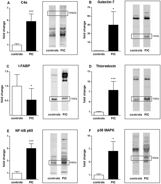
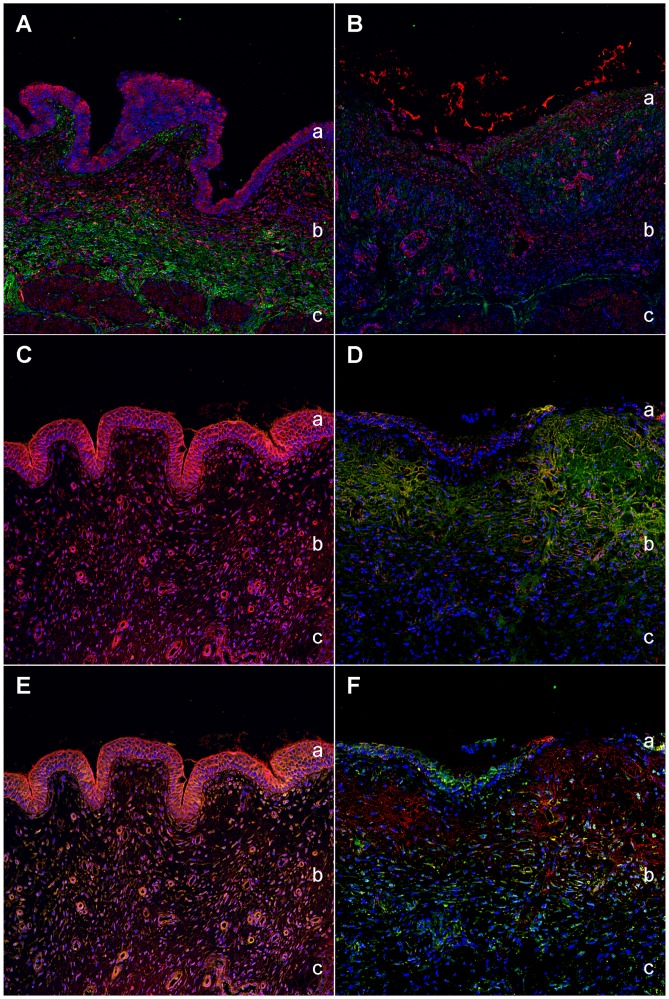
Feline idiopathic cystitis (FIC) is the only spontaneous animal model for human interstitial cystitis (IC), as both possess a distinctive chronical and relapsing character. Underlying pathomechanisms of both diseases are not clearly established yet. We recently detected increased urine fibronectin levels in FIC cases. The purpose of this study was to gain further insight into the pathogenesis by assessing interacting partners of fibronectin in urine of FIC affected cats. Several candidate proteins were identified via immunoprecipitation and mass spectrometry. Considerable changes in FIC conditions compared to physiological expression of co-purified proteins were detected by Western blot and immunohistochemistry. Compared to controls, complement C4a and thioredoxin were present in higher levels in urine of FIC patients whereas loss of signal intensity was detected in FIC affected tissue. Galectin-7 was exclusively detected in urine of FIC cats, pointing to an important role of this molecule in FIC pathogenesis. Moderate physiological signal intensity of galectin-7 in transitional epithelium shifted to distinct expression in transitional epithelium under pathophysiological conditions. I-FABP expression was reduced in urine and urinary bladder tissue of FIC cats. Additionally, transduction molecules of thioredoxin, NF-κB p65 and p38 MAPK, were examined. In FIC affected tissue, colocalization of thioredoxin and NF-κB p65 could be demonstrated compared to absent coexpression of thioredoxin and p38 MAPK. These considerable changes in expression level and pattern point to an important role for co-purified proteins of fibronectin and thioredoxin-regulated signal transduction pathways in FIC pathogenesis. These results could provide a promising starting point for novel therapeutic approaches in the future.


Conflict of interest statement

Competing Interests: The authors have declared that no competing interests exist.

Figures

Figure 1
Figure 1. Quantification of signal intensities of co-purified candidate proteins of healthy and FIC diseased cases.
Western blot signal intensity of healthy controls (white columns, left, n = 20) and diseased urine samples (grey columns, right, n = 16) were compared for the following co-purified proteins: Complement component 4a (C4a) (A), galectin-7 (B), fatty acid-binding protein, intestinal (I-FABP) (C), thioredoxin (D), NF-κB p65 (E) and p38 MAPK (F). The according Western blot strips visualize the quantitative difference of signal intensities. The left strips show representative control blots, the right ones blots with FIC urine. The corresponding band sizes are displayed in black boxes. Signals were quantified by densitometry and statistical significance was calculated using the Mann-Whitney test. Data are represented in a column bar graph as means with SEM. Expression level of C4a (A) shows a significant (***p≤0.001) increase in urine of FIC diseased cases with an almost 5-fold higher concentration compared to urine of healthy controls. Galectin-7 (B) is significantly (*p≤0.05) upregulated in FIC diseased samples compared to healthy control samples with a 45-fold higher expression in FIC affected cases. Quantification of I-FABP expression (C) results in a significant (*p≤0.05) decrease in urine of FIC cases compared to urine of healthy controls. Thioredoxin (D) is significantly (***p≤0.001) increased by a factor of 7.5 in FIC compared to urine of controls. Abundances of NF-kB p65 (E) significantly (***p≤0.001) increase in diseased specimens compared to healthy specimens with a 5-fold higher expression just as p38 MAPK (F) showing an 6.5-fold higher expression (*p≤0.05) in FIC diseased urines in comparison to healthy control urines.
Figure 2
Figure 2. H&E staining of normal feline bladder (A) and FIC diseased bladder (B).
Histological sections stained with Haematoxylin and Eosin. Healthy urinary bladder section (A) shows characteristic architecture compared with FIC diseased bladder tissue (B), where a loss of normal bladder wall physiology can be seen. Notice the marked loss of transitional cell epithelium, the intramucosal bleeding and oedema in the FIC section (B). a = Transitional cell epithelium, b = Lamina propria mucosae, c = Loss of transitional cell epithelium, d = Intramucosal bleeding and oedema.
Figure 3
Figure 3. Expression of co-purified proteins in healthy bladder tissue.
Immunohistochemical double labelling of CD117 and co-purified proteins in a representative healthy bladder tissue. DIC image of healthy bladder tissue (A). CD117 (green) shows a marked reactivity in the epithelial cells of the urothel and in the interstitial cells of the lamina propria of the healthy bladder (B). Overlay image of C4a (red) and CD117 (green) reveals considerable co-localization (overlapping results in yellow colour) at the cell nuclei of the urothelial cells and a scattered expression in the interstitial cells of the lamina propria (C). Galectin-7 (red) and CD117 (green) show a co-localization in the umbrella cells (marked with an asterisk) of the transitional cell epithelium, whereas reactivity of both proteins in the epithelial cells of the urothel indicate a co-expression. Cells of the lamina propria are only CD117 positive (D). I-FABP (red) and CD117 (green) overlay is visible only in the interstitial cells of the lamina propria. Additionally, I-FABP reactivity is seen extracellularly and is distinctly expressed in the basal membrane (E). Thioredoxin (red) and CD117 (green) co-localize distinctly at all cell nuclei of the transitional epithelial cells and in the interstitial cells of the subepithelial tunic (F). The blue colour reveals staining of cell nuclei (DAPI). a = Transitional cell epithelium, b = Lamina propria mucosae, c = Inserted box shows magnification of respective cells in the lamina propria mucosae.
Figure 4
Figure 4. Expression pattern of fibronectin and its co-purified proteins in healthy and diseased bladder tissue.
Urinary bladder expression of fibronectin (green) and its co-purified proteins (red) in a representative healthy (left panels) and FIC diseased bladder (right panels). Physiological distribution of fibronectin (green) in healthy bladder (A). Extracellular matrix of the lamina propria mucosae and the muscle tunic show a distinct immunoreactivity for fibronectin, whereas a loss in FIC affected bladder tissue (B), especially in the subepithelial and muscular tunices, is evident. C4a (red) is moderately expressed in the apical transitional cell epithelium of the physiological bladder (C) and disappears in the FIC affected bladder tissue (D). Galectin-7 (red) is expressed especially in umbrella cells of the transitional epithelium and around blood vessels in the lamina propria under normal condition (E). In contrast, expression changes profoundly in FIC affected bladders to distinct expression in the transitional cell epithelium (F). Reactivity of I-FABP (red) throughout all tunices in healthy bladder (G) almost disappears in FIC diseased tissue (H). Thioredoxin (red) reveals a predominant signal in the entire healthy bladder tissue (I) compared to a leakage of thioredoxin into the lumen (arrow) of FIC affected bladder tissue resulting in a slight immunoreactivity of the diseased bladder tissue (J). The blue colour reveals staining of cell nuclei (DAPI). a = Transitional cell epithelium, b = Lamina propria mucosae, c = Muscle tunic, d = Normal vessel.
Figure 5
Figure 5. Immunohistochemical double labelling of candidates in healthy (left panels) and diseased bladder tissue (right panels).
Immunohistochemical double staining of a healthy urinary bladder (A) shows considerable colocalization of fibronectin (green) and its interactor thioredoxin (red) in the subepithelial and muscular tunices. In contrast, lack of green and red colour is evident in FIC (B), indicating a loss of both fibronectin and thioredoxin from its normal distribution in healthy bladder tissue. Overlay image of thioredoxin (red) and NF-κB p65 (green) in a healthy bladder tissue (C) shows a predominant signal of thioredoxin in all tunices of the bladder, whereas NF-κB p65 is not detectable in any tunic of the healthy bladder. In contrast, thioredoxin and NF-κB p65 colocalize (overlapping results in yellow colour) in the lamina propria mucosae with the highest expression in the extracellular matrix of FIC diseased bladder tissue (D). P38 MAPK signal is of moderate intensity localized in transitional epithelium cells and around few blood vessels in the healthy bladder tissue (E). Note that the signal is exclusively of yellow colour indicating a colocalization with thioredoxin, whereas a green colour signal is not visible at all. In contrast, p38 MAPK (green) was highly expressed in the cytoplasm of umbrella cells of the transitional cell epithelium as well as a scattered expression around cell nuclei in the subepithelial and muscular tunices of FIC diseased bladder sections without distinct colocalization (F). The blue colour reveals staining of cell nuclei (DAPI). a = Transitional cell epithelium, b = Lamina propria mucosae, c = Muscle tunic.

Similar articles

Cited by

References

    1. Westropp JL, Buffington CA (2002) In vivo models of interstitial cystitis. J Urol 167: 694–702. - PubMed
    1. Gunn-Moore DA (2003) Feline lower urinary tract disease. J Feline Med Surg 5: 133–138. - PMC - PubMed
    1. Lavelle JP, Meyers SA, Ruiz WG, Buffington CA, Zeidel ML, et al. (2000) Urothelial pathophysiological changes in feline interstitial cystitis: a human model. Am J Physiol Renal Physiol 278: F540–553. - PubMed
    1. Buffington CA, Chew DJ, Woodworth BE (1999) Feline interstitial cystitis. J Am Vet Med Assoc 215: 682–687. - PubMed
    1. Nordling J, Fall M, Hanno P (2011) Global concepts of bladder pain syndrome (interstitial cystitis). World J Urol. - PubMed

Publication types

MeSH terms