. 2012;7(12):e51433.
doi: 10.1371/journal.pone.0051433. Epub 2012 Dec 6.

Neto2 interacts with the scaffolding protein GRIP and regulates synaptic abundance of kainate receptors

Affiliations

Neto2 interacts with the scaffolding protein GRIP and regulates synaptic abundance of kainate receptors

Man Tang et al. PLoS One. 2012.

Abstract

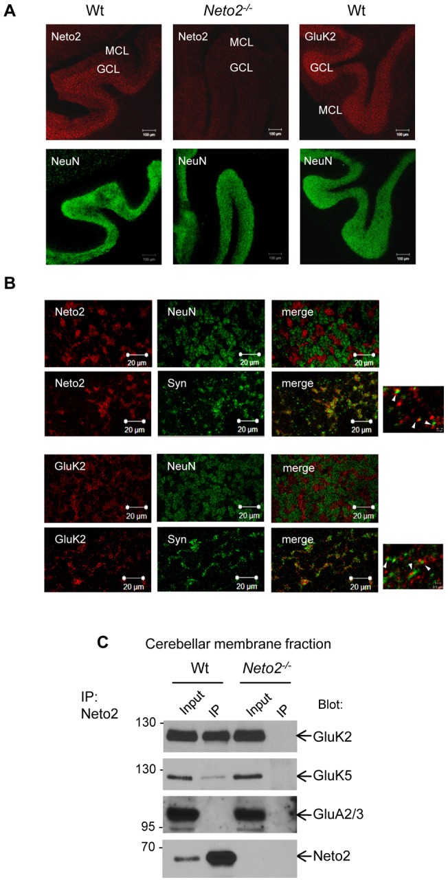
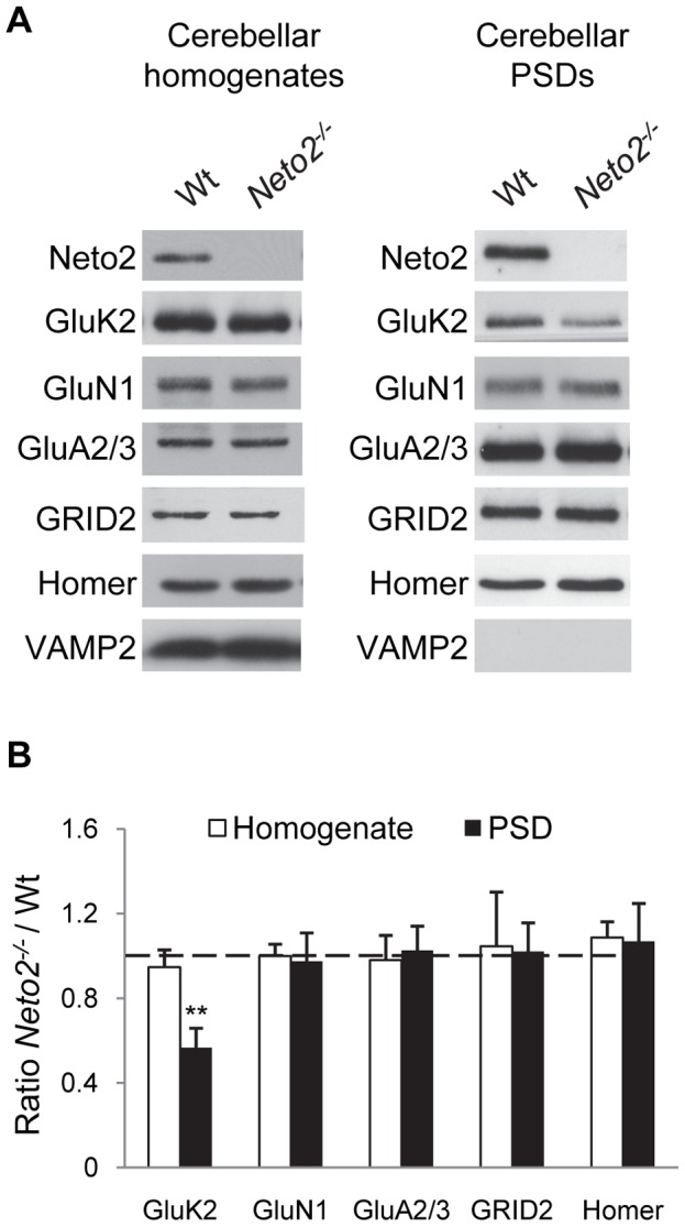
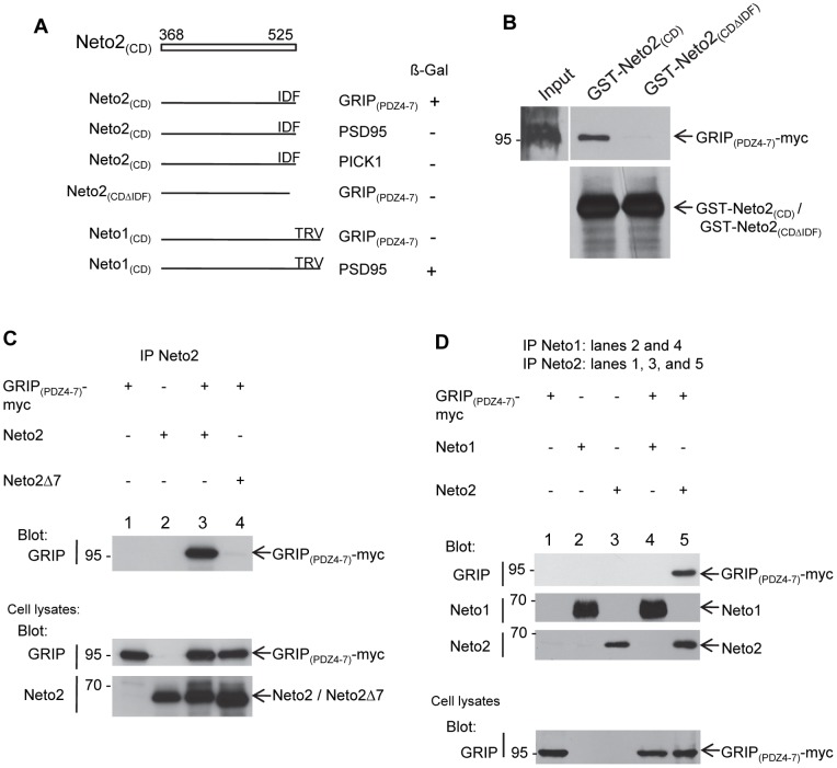
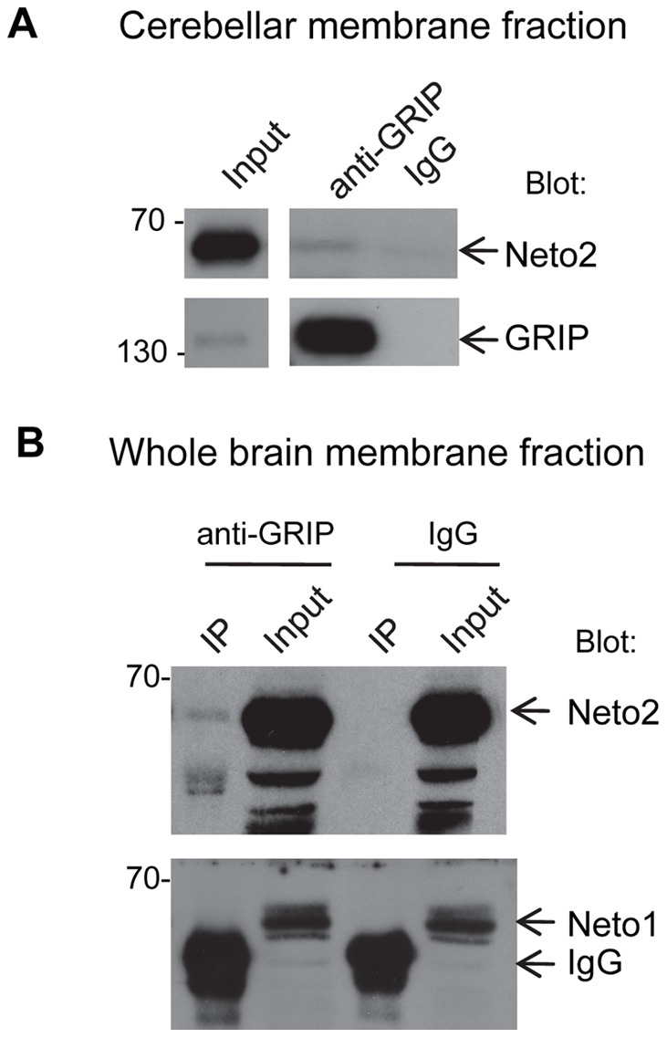
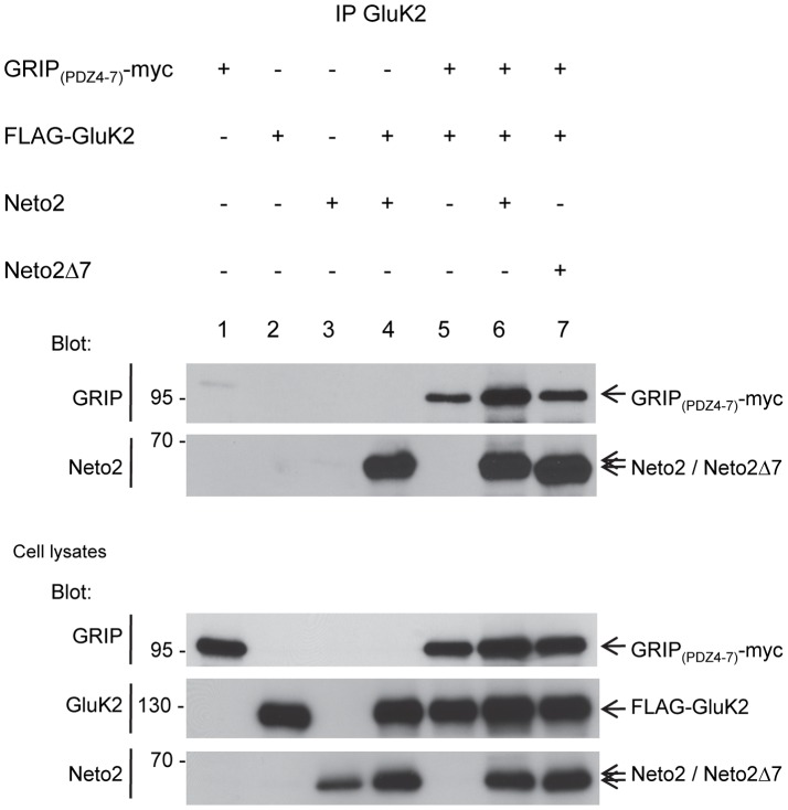
Kainate receptors (KARs) are a class of ionotropic glutamate receptors that are expressed throughout the central nervous system. The function and subcellular localization of KARs are tightly regulated by accessory proteins. We have previously identified the single-pass transmembrane proteins, Neto1 and Neto2, to be associated with native KARs. In the hippocampus, Neto1, but not Neto2, controls the abundance and modulates the kinetics of postsynaptic KARs. Here we evaluated whether Neto2 regulates synaptic KAR levels in the cerebellum where Neto1 expression is limited to the deep cerebellar nuclei. In the cerebellum, where Neto2 is present abundantly, we found a ~40% decrease in GluK2-KARs at the postsynaptic density (PSD) of Neto2-null mice. No change, however, was observed in total level of GluK2-KARs, thereby suggesting a critical role of Neto2 on the synaptic localization of cerebellar KARs. The presence of a putative class II PDZ binding motif on Neto2 led us to also investigate whether it interacts with PDZ domain-containing proteins previously implicated in regulating synaptic abundance of KARs. We identified a PDZ-dependent interaction between Neto2 and the scaffolding protein GRIP. Furthermore, coexpression of Neto2 significantly increased the amount of GRIP associated with GluK2, suggesting that Neto2 may promote and/or stabilize GluK2:GRIP interactions. Our results demonstrate that Neto2, like Neto1, is an important auxiliary protein for modulating the synaptic levels of KARs. Moreover, we propose that the interactions of Neto1/2 with various scaffolding proteins is a critical mechanism by which KARs are stabilized at diverse synapses.


Conflict of interest statement

Competing Interests: The authors have declared that no competing interests exist.

Figures

Figure 1
Figure 1. Neto2 is associated with KARs in the cerebellum.
(A) Confocal micrographs of immunostained cerebellar slices. Antibodies used for immunostaining are indicated in the top left corner of each image. In the cerebellum, the NeuN antibody stains the neuronal nuclei of granule cells but does not recognize Purkinje cells. MCL, molecular cell layer; GCL, granule cell layer; Wt, wild-type sections; Neto2−/−, Neto2-null sections. Scale bar, 100 μm. (B) High-magnification confocal microscopy of the cerebellar granule cell layer immunostained with Neto2, GluK2, NeuN, or synaptophysin antibodies. Scale bar, 20 μm; scale bar (small panels on the right), 5 μm (C) Immunoblot of immunoprecipitates from the cerebellum. Blot: antibody used for immunoblot analysis; IP: immunoprecipitate. The input represents 2% of the material used in the immunoprecipitation experiment.
Figure 2
Figure 2. KARs are reduced in the cerebellar PSD of Neto2-null mice.
(A) Immunoblots (representative of three experiments) of proteins from cerebellar homogenates and cerebellar PSD fractions of wild-type (Wt) and Neto2-null (Neto2−/−) mice. Antibodies used for detection are indicated on the left. (B) Histogram showing normalized levels of different proteins in Neto2-null cerebellar homogenates relative to that of wild-type (white bars), and in Neto2-null cerebellar PSD fractions relative to that of wild-type (black bars); **, p<0.01, paired t-test, n = 3.
Figure 3
Figure 3. Neto2 interacts with GRIP through a C-terminal binding motif.
(A) Yeast two-hybrid analysis of Neto2 interaction with PDZ domain-containing proteins. ß-gal, ß-galactosidase assay. Numbers on top of the white bar indicate the amino acid residues corresponding to the Neto2 cytoplasmic domain. Neto2(CD), cytoplasmic domain of Neto2; Neto2(CDΔIDF), cytoplasmic domain of Neto2 lacking the last three C-terminal amino acids (IDF); Neto1(CD), cytoplasmic domain of Neto1 encompassing amino acids 345–533. The last three C-terminal amino acids of Neto1 are TRV. (B) Pull-down of recombinant GRIP(PDZ4–7)-myc with GST fused to the cytoplasmic domain of Neto2 (GST-Neto2(CD)) or with GST fused to the deletion mutant ΔIDF (GST-Neto2(CDΔIDF)). (C, D) Immunoblots of immunoprecipitates from transfected COS-7 cell lysates. The cDNAs used for transfection are shown above each lane. Neto2Δ7, Neto2 lacking the last seven C-terminal residues; blot, antibody used for immunoblot analysis; IP, antibody used for immunoprecipitation.
Figure 4
Figure 4. Neto2 associates with GRIP in vivo.
(A, B) Immunoblots of immunoprecipitates from cerebellar (A) or whole brain (B) membrane fractions. Samples were subjected to immunoprecipitation with an anti-GRIP antibody, or with normal rabbit IgGs (IgG), as the negative control. Blot, antibody used for immunoblot analysis; IP, immunoprecipitate.
Figure 5
Figure 5. Neto2 increases the interaction between GRIP and GluK2.
Immunoblot of immunoprecipitates from lysates of transfected COS-7 cells. The cDNAs used for transfection are shown above each lane. Neto2Δ7, Neto2 lacking the last seven C-terminal residues. Blot, antibody used for immunoblot analysis; IP, antibody used for immunoprecipitation.

References

    1. Contractor A, Mulle C, Swanson GT (2011) Kainate receptors coming of age: milestones of two decades of research. Trends Neurosci 34: 154–163. - PMC - PubMed
    1. Pinheiro PS, Mulle C (2008) Presynaptic glutamate receptors: physiological functions and mechanisms of action. Nat Rev Neurosci 9: 423–436. - PubMed
    1. Castillo PE, Malenka RC, Nicoll RA (1997) Kainate receptors mediate a slow postsynaptic current in hippocampal CA3 neurons. Nature 388: 182–186. - PubMed
    1. Vignes M, Collingridge GL (1997) The synaptic activation of kainate receptors. Nature 388: 179–182. - PubMed
    1. Cossart R, Esclapez M, Hirsch JC, Bernard C, Ben-Ari Y (1998) GluR5 kainate receptor activation in interneurons increases tonic inhibition of pyramidal cells. Nat Neurosci 1: 470–478. - PubMed

Publication types