. 2012;7(12):e51477.
doi: 10.1371/journal.pone.0051477. Epub 2012 Dec 7.

Insights into factorless translational initiation by the tRNA-like pseudoknot domain of a viral IRES

Affiliations

Insights into factorless translational initiation by the tRNA-like pseudoknot domain of a viral IRES

Hilda H T Au et al. PLoS One. 2012.

Abstract

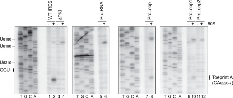
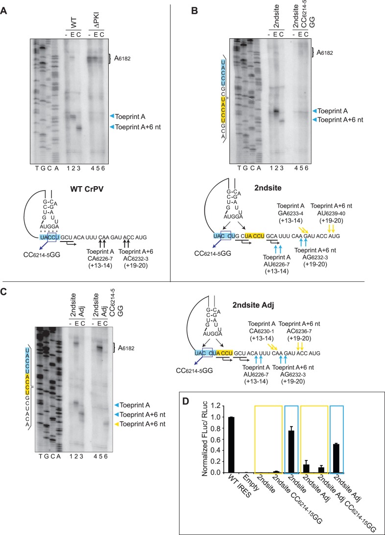
The intergenic region internal ribosome entry site (IGR IRES) of the Dicistroviridae family adopts an overlapping triple pseudoknot structure to directly recruit the 80S ribosome in the absence of initiation factors. The pseudoknot I (PKI) domain of the IRES mimics a tRNA-like codon:anticodon interaction in the ribosomal P site to direct translation initiation from a non-AUG initiation codon in the A site. In this study, we have performed a comprehensive mutational analysis of this region to delineate the molecular parameters that drive IRES translation. We demonstrate that IRES-mediated translation can initiate at an alternate adjacent and overlapping start site, provided that basepairing interactions within PKI remain intact. Consistent with this, IGR IRES translation tolerates increases in the variable loop region that connects the anticodon- and codon-like elements within the PKI domain, as IRES activity remains relatively robust up to a 4-nucleotide insertion in this region. Finally, elements from an authentic tRNA anticodon stem-loop can functionally supplant corresponding regions within PKI. These results verify the importance of the codon:anticodon interaction of the PKI domain and further define the specific elements within the tRNA-like domain that contribute to optimal initiator Met-tRNA(i)-independent IRES translation.


Conflict of interest statement

Competing Interests: Author Eric Jan is an Academic Editor for PLOS ONE. This does not alter the authors' adherence to all the PLOS ONE policies on sharing data and materials.

Figures

Figure 1
Figure 1. Chimeric IRESs containing prolyl-tRNA and wild-type CrPV PKI regions.
(A) Top. Schematic of the authentic prolyl-tRNA and wild-type CrPV IRES. The expected location of the positioning toeprint (Toeprint A) is indicated. Bottom. Schematic of the dicistronic reporter constructs containing regions of the IRES PKI domain that have been substituted by prolyl-tRNA elements (grey) or bearing mutations in PKI (black box) (B) Translational activities of chimeric and mutant IRESs. Top. Uncapped dicistronic reporter RNAs used in translation and toeprinting/primer extension assays. The upstream renilla luciferase (RLuc) and downstream firefly luciferase (FLuc) cistrons are expressed by scanning-mediated and IRES-mediated translation, respectively. Bottom. Uncapped dicistronic reporter RNAs containing the wild-type or mutant IGR IRES in the intercistronic space were incubated in RRL in the presence of [35S]-methionine. Shown are the average values ± S.D. for the ratios of FLuc to RLuc expression, normalized to the wild-type IGR IRES from at least three independent experiments. (C) Summary of the translational activities and toeprint intensities (from Figure 2) for the chimeric and mutant IRESs. Toeprint intensities were measured as a fraction of the radioactive counts for Toeprint A over the total radioactive counts within each lane, normalized to that of the wild-type IGR IRES.
Figure 2
Figure 2. Toeprinting analysis of chimeric IRESs.
Dicistronic reporter RNAs containing the wild-type or mutant IGR IRES were incubated in the absence [−] or presence [+] of purified salt-washed HeLa 80S (100 nM). Primer extension analysis was performed using PrEJ69 in the presence of α-[32P]-dATP. Reaction products were resolved by denaturing polyacrylamide gel electrophoresis and visualized by autoradiography. The sequencing reactions for each construct are shown on the left, with the respective nucleotide numbers as indicated. The locations of the major toeprints are as indicated on the right.
Figure 3
Figure 3. The IRES helical stem tolerates bulges and a 1-basepair deletion.
(A) Schematic of the IRES PKI helical stem is shown, with the region bearing mutations highlighted in grey. The respective mutations resulting in a 1-nucleotide bulge (ΔU6176), 2-nucleotide bulge (ΔUU6176-7), 1-basepair deletion (ΔU6176/A6200) and 2-basepair deletion (ΔUU6176-7/AA6199-200) are shown. (B) Translational activities of mutants bearing alterations in the PKI helical stem. Uncapped dicistronic reporter RNAs containing the wild-type or mutant IGR IRES in the intercistronic space were incubated in RRL in the presence of [35S]-methionine. Shown are the average values ± S.D. for the ratios of FLuc to RLuc expression, normalized to the wild-type IGR IRES from at least three independent experiments. (C) Toeprinting analysis for IRESs bearing mutations in the helical stem. Dicistronic reporter RNAs containing the wild-type or mutant IGR IRES were incubated in the absence [−] or presence [+] of purified salt-washed HeLa 80S (100 nM). Primer extension analysis was performed using PrEJ69 in the presence of α-[32P]-dATP. Reaction products were resolved by denaturing polyacrylamide gel electrophoresis and visualized by autoradiography. The sequencing reaction for the wild-type IRES is shown on the left, with the respective nucleotide numbers as indicated. The locations of the major toeprints are as indicated on the right. (D) Summary of the translational activities and toeprint intensities for the mutant IRESs. Toeprint intensities were measured as a fraction of the radioactive counts for Toeprint A over the total radioactive counts within each lane, normalized to that of the wild-type IGR IRES.
Figure 4
Figure 4. Alterations in the length of the variable loop region (VLR) adversely affect IRES-mediated translation.
(A) The schematic of the CrPV IGR IRES secondary structure is shown. The nucleotides constituting the VLR are highlighted in grey. The expected locations of a properly positioned ribosome on the IGR IRES (Toeprint A) and a translocated ribosome (Toeprint A+6 nt) are indicated. The first nucleotide of the codon occupying the ribosomal A site is indicated (+1). (B) Translational activities of VLR insertion/deletion mutants. Uncapped dicistronic reporter RNAs containing the wild-type or mutant IGR IRES in the intercistronic space were incubated in RRL in the presence of [35S]-methionine. Shown are the average values ± S.D. for the ratios of FLuc to RLuc expression, normalized to the wild-type IGR IRES from at least three independent experiments. (C) Summary of IRES translational activities and toeprint intensities in RRL for each construct. Insertion/deletion mutations in the VLR are shown, with inserted and deleted nucleotides highlighted in grey and denoted with dash lines, respectively. Deviations from the wild-type length for each construct are indicated. Toeprint intensities were measured as a fraction of the radioactive counts for Toeprint A or Toeprint A+6 nt over the total radioactive counts within each lane, normalized to that of the wild-type IGR IRES (from Figure 5A).
Figure 5
Figure 5. Toeprinting analysis of VLR insertion/deletion mutant IGR IRESs.
Dicistronic reporter RNAs containing the wild-type or mutant IGR IRES were incubated in (A) RRL without drug treatment [−], or under treatment with edeine [E] or cycloheximide [C] or (B) in the absence [−] or presence [+] of purified salt-washed HeLa 80S ribosomes (100 nM) as described in the Materials and Methods section. Primer extension analysis was performed using PrEJ69 in the presence of α- [32P]-dATP. Reaction products were resolved by denaturing polyacrylamide gel electrophoresis and visualized by autoradiography. The sequencing reactions for the wild-type IRES are shown on the left, with the respective nucleotide numbers as indicated. The locations of the major toeprints are as indicated on the right. Shown are representative gels from at least two independent experiments.
Figure 6
Figure 6. The conserved A6205 is important for IRES-mediated translation.
(A) Sequence alignment of the variable loop region (VLR) of members of the type I IGR IRESs. Nucleotides involved in basepairing of the helical stem are bolded and those comprising pseudoknot I are highlighted in grey. The nucleotides constituting the VLR are denoted, with conserved positions highlighted in red. The assignments are made based on the secondary structure predictions of the IGR IRESs. The translational start site is indicated. (B) The schematic of the PKI domain is shown with the nucleotides constituting the VLR highlighted in grey. Four conserved nucleotides within the VLR, A6205, A6207, A6208 and U6211 are denoted by boxes and were mutated to each of the alternate Watson-Crick bases. (C) Translational activities of VLR point mutants. Uncapped dicistronic reporter RNAs containing the wild-type or mutant IGR IRES in the intercistronic space were incubated in RRL in the presence of [35S]-methionine. Shown are the average values ± S.D. for the ratios of FLuc to RLuc expression, normalized to the wild-type IGR IRES from at least three independent experiments. (D) Toeprinting/primer extension assay of VLR point mutants. Dicistronic reporter RNAs containing the wild-type or mutant IGR IRES were incubated in RRL under treatment with edeine [E] or cycloheximide [C] as described in the Materials and Methods section. Primer extension analysis was performed using PrEJ69 in the presence of α- [32P]-dATP. Reaction products were resolved by denaturing polyacrylamide gel electrophoresis and visualized by autoradiography. The sequencing reactions for the wild-type IRES are shown on the left, with the respective nucleotide numbers as indicated. The locations of the major toeprints are as indicated on the right. Shown is a representative gel from at least two independent experiments.
Figure 7
Figure 7. IGR IRES-mediated translation initiation at an alternate translational start site.
Schematic of the construct of the wild-type IGR IRES PKI domain (A, bottom) and constructs which contain an alternate translational start site 2 nucleotides downstream [2ndsite] (B, bottom), or adjacent to and overlapping with the authentic start site [2ndsite Adj] (C, right). The authentic and inserted translation start sites are highlighted in blue and yellow, respectively. The expected locations of a properly positioned ribosome (Toeprint A) and a translocated ribosome (Toeprint A+6 nt) from the authentic and inserted sites are denoted by arrows of the corresponding colors for each construct. (A and B (Top), C, (left)) Toeprinting analysis for the respective constructs. Dicistronic reporter RNAs containing the wild-type or mutant IGR IRES were incubated in RRL without drug treatment [−], or under treatment with edeine [E] or cycloheximide [C] as described in the Materials and Methods section. Primer extension analysis was performed using PrEJ69 in the presence of α- [32P] dATP. Reaction products were resolved by denaturing polyacrylamide gel electrophoresis and visualized by autoradiography. The sequencing reactions for the constructs are shown, with the nucleotides corresponding to the authentic and inserted sites highlighted in blue and yellow, respectively. The locations of the major toeprints are as indicated to the right of the gel in the same color. (D) IRES translational activities of reporter constructs containing alternate translational start sites. Uncapped dicistronic reporter RNAs containing the wild-type or mutant IGR IRES in the intercistronic space were incubated in RRL in the presence of [35S]-methionine. Shown are the ratios of FLuc to RLuc expression, normalized to the wild-type IGR IRES. Translational activities monitored in the reading frame of the authentic and inserted start sites are highlighted in blue and yellow, respectively. Shown are average values from at least three independent experiments ± S.D.

References

    1. Pestova TV, Lorsch JR, Hellen CU (2007) The Mechanism of Translation Initiation in Eukaryotes. In: Mathews MB, Sonenberg N, Hershey J, editors. Translational Control in Biology and Medicine. Cold Spring Harbor, NY: Cold Spring Harbor Laboratory Press. 87.
    1. Hellen CU, Sarnow P (2001) Internal ribosome entry sites in eukaryotic mRNA molecules. Genes Dev 15: 1593–1612. - PubMed
    1. Doudna JA, Sarnow P (2007) Translation Initiation by Viral Internal Ribosome Entry Sites. In: Mathews MB, Sonenberg N, Hershey J, editors. Translation Control in Biology and Medicine. Cold Spring Harbor, NY: Cold Spring Harbor Laboratory Press. 129.
    1. Kieft JS (2008) Viral IRES RNA structures and ribosome interactions. Trends Biochem Sci 33: 274–283. - PMC - PubMed
    1. Pestova TV, Shatsky IN, Fletcher SP, Jackson RJ, Hellen CU (1998) A prokaryotic-like mode of cytoplasmic eukaryotic ribosome binding to the initiation codon during internal translation initiation of hepatitis C and classical swine fever virus RNAs. Genes Dev 12: 67–83. - PMC - PubMed

Publication types