. 2012;6(12):e1923.
doi: 10.1371/journal.pntd.0001923. Epub 2012 Dec 6.

Cathepsin B & L are not required for ebola virus replication

Affiliations

Cathepsin B & L are not required for ebola virus replication

Andrea Marzi et al. PLoS Negl Trop Dis. 2012.

Abstract

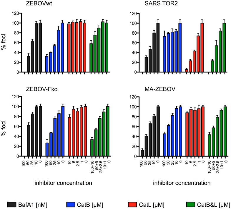
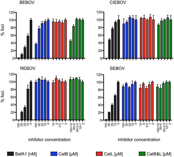
Ebola virus (EBOV), family Filoviridae, emerged in 1976 on the African continent. Since then it caused several outbreaks of viral hemorrhagic fever in humans with case fatality rates up to 90% and remains a serious Public Health concern and biothreat pathogen. The most pathogenic and best-studied species is Zaire ebolavirus (ZEBOV). EBOV encodes one viral surface glycoprotein (GP), which is essential for replication, a determinant of pathogenicity and an important immunogen. GP mediates viral entry through interaction with cellular surface molecules, which results in the uptake of virus particles via macropinocytosis. Later in this pathway endosomal acidification activates the cysteine proteases Cathepsin B and L (CatB, CatL), which have been shown to cleave ZEBOV-GP leading to subsequent exposure of the putative receptor-binding and fusion domain and productive infection. We studied the effect of CatB and CatL on in vitro and in vivo replication of EBOV. Similar to previous findings, our results show an effect of CatB, but not CatL, on ZEBOV entry into cultured cells. Interestingly, cell entry by other EBOV species (Bundibugyo, Côte d'Ivoire, Reston and Sudan ebolavirus) was independent of CatB or CatL as was EBOV replication in general. To investigate whether CatB and CatL have a role in vivo during infection, we utilized the mouse model for ZEBOV. Wild-type (control), catB(-/-) and catL(-/-) mice were equally susceptible to lethal challenge with mouse-adapted ZEBOV with no difference in virus replication and time to death. In conclusion, our results show that CatB and CatL activity is not required for EBOV replication. Furthermore, EBOV glycoprotein cleavage seems to be mediated by an array of proteases making targeted therapeutic approaches difficult.


Conflict of interest statement

The authors have declared that no competing interests exist.

Figures

Figure 1
Figure 1. Zaire ebolavirus entry is CatB mediated.
Vero E6 cells were treated prior to infection for one hour with inhibitors directed against the indicated cathepsin(s) or endosomal acidification (BafA1). Following infection the cells were washed and a carboxymethyl cellulose overlay was added containing inhibitor (BafA1 100, 50, 25, 10 nM; CA-074 (CatB) 100, 50, 25, 10 µM; CatL inhibitor V 10, 5, 2.5, 1 µM). SARS-CoV infected cells were fixed on day 3, stained with crystal violet and plaques were counted. ZEBOV infected cells were fixed on day 4, foci were stained with an antibody against ZEBOV-VP40 and counted. The number of foci and plaques without inhibitor was set as 100%. A representative experiment performed in triplicates is shown. Error bars indicate the standard error of the mean. ZEBOVwt = Zaire ebolavirus, strain Mayinga; ZEBOV-Fko = Zaire ebolavirus furin cleavage site knockout mutant; MA-ZEBOV = mouse-adapted Zaire ebolavirus.
Figure 2
Figure 2. Entry of other Ebola viruses is CatB-independent.
Vero E6 cells were treated prior to infection for one hour with inhibitors directed against the indicated cathepsin(s) or endosomal acidification (BafA1). Following infection the cells were washed and a carboxymethyl cellulose overlay containing inhibitor (BafA1 100, 50, 25, 10 nM; CA-074 (CatB) 100, 50, 25, 10 µM; CatL inhibitor V 10, 5, 2.5, 1 µM) was added. After fixation on day 4, foci were stained with antibodies against VP40 (BEBOV = Bundibugyo ebolavirus, CIEBOV = Côte d'Ivoire ebolavirus, SEBOV = Sudan ebolavirus, strain Boniface) or NP (REBOV = Reston ebolavirus, strain Pennsylvania) and counted. The number of foci without inhibitor was set as 100%. A representative experiment performed in triplicates is shown. Error bars indicate the standard error of the mean.
Figure 3
Figure 3. Zaire ebolavirus growth is CatB and CatL-independent.
(A) Vero E6 cells were seeded the night before infection in a 24-well-plate. One hour prior to infection cells were incubated with inhibitors directed against the indicated cathepsin(s) or endosomal acidification (Baf A1). ZEBOVwt was added at an MOI of 1 and incubated for one hour at 37°C. After three washes the cells were covered with 1 ml medium containing 50 nM BafA1, 50 µM CA-074, 5 µM CatL inhibitor V or no inhibitor and incubated for 4 days. (B) and (C) MEF cell lines were seeded the night before infection in a 24-well-plate. Cells were infected for 1 hour with 0.2 ml ZEBOVwt (B) or MA-ZEBOV (C) at a MOI of 1. After three washes the cells were covered with 1 ml medium and incubated for 4 days. For all experiments, samples were collected at 0, 12, 24, 48, 72 and 96 hours post infection and infectious titers were determined. A representative experiment performed in triplicates is shown. Error bars indicate the standard error of the mean.
Figure 4
Figure 4. CatB−/− and catL−/− mice succumb to Ebola virus but not to VSV infection.
Groups of mice were i.p. infected with 10 ffu MA-ZEBOV (1,000 LD50) or 1×10⁵ pfu VSV (serotype Indiana) and monitored daily for weight loss and other signs of illness. Survival (A) and weight curves (B) for MA-ZEBOV infection are shown. Body weights of VSV-infected mice are shown in (C). VSV antibodies were detected using ELISA to confirm infection (D).
Figure 5
Figure 5. Zaire ebolavirus replicates to similar titers in knockout and control mice.
CatB−/−, catL−/− or control mice (n = 3) were i.p. infected with 1,000 LD50 of MA-ZEBOV and euthanized at the indicated time point. Liver, spleen and blood samples were taken on day 3 (A) and day 7 (B) and viral titers were determined. A single 50% tissue culture infectious dose (TCID50) value is depicted for each mouse. Bars indicate the mean value.

References

    1. Sanchez A, Geisbert TW, Feldmann H (2006) Filoviridae: Marburg and Ebola viruses Fields Virology 1409–1448.
    1. Towner JS, Sealy TK, Khristova ML, Albarino CG, Conlan S, et al. (2008) Newly discovered ebola virus associated with hemorrhagic fever outbreak in Uganda. PLoS Pathog 4: e1000212. - PMC - PubMed
    1. Takada A (2012) Filovirus tropism: cellular molecules for viral entry. Frontiers in Microbiology 3/00034. - PMC - PubMed
    1. Carette JE, Raaben M, Wong AC, Herbert AS, Obernosterer G, et al. (2011) Ebola virus entry requires the cholesterol transporter Niemann-Pick C1. Nature 477: 340–343. - PMC - PubMed
    1. Cote M, Misasi J, Ren T, Bruchez A, Lee K, et al. (2011) Small molecule inhibitors reveal Niemann-Pick C1 is essential for Ebola virus infection. Nature 477: 344–348. - PMC - PubMed

Publication types

MeSH terms