

Clofazimine modulates the expression of lipid metabolism proteins in Mycobacterium leprae-infected macrophages
- PMID: 23236531
- PMCID: PMC3516583
- DOI: 10.1371/journal.pntd.0001936
Clofazimine modulates the expression of lipid metabolism proteins in Mycobacterium leprae-infected macrophages
Abstract
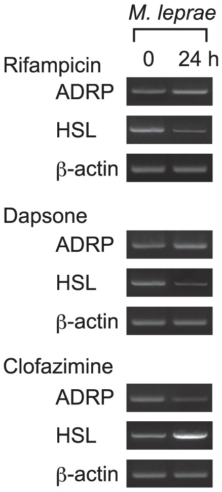
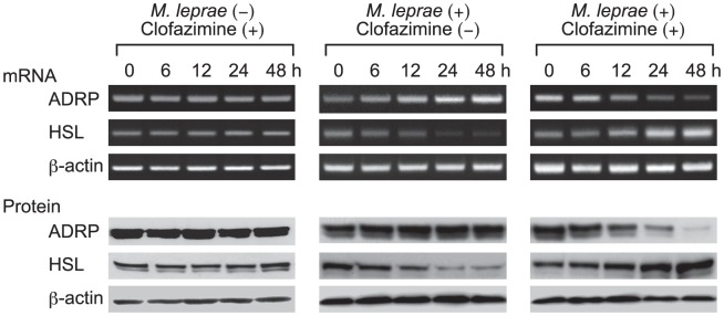
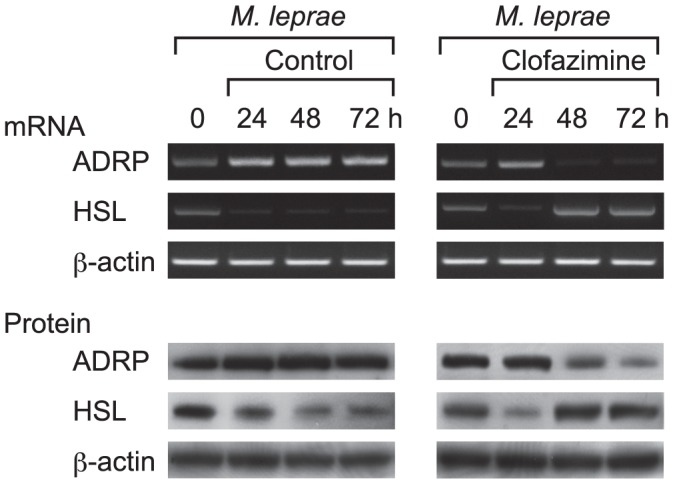
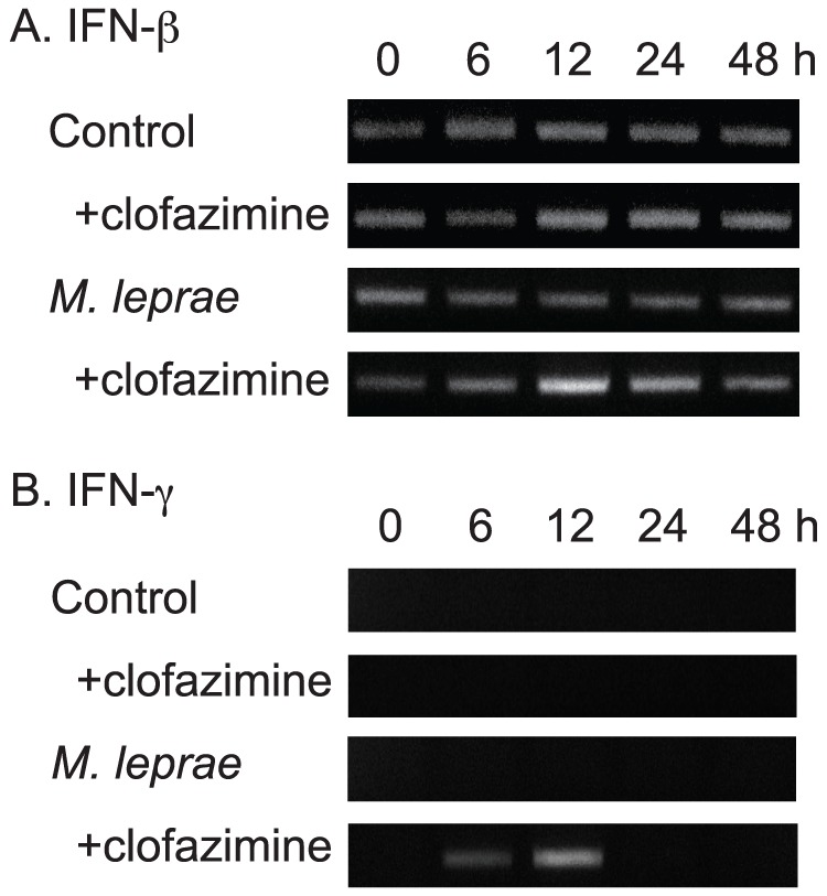
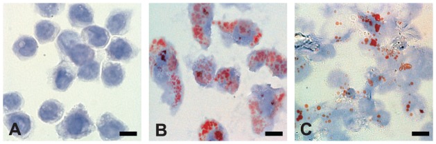
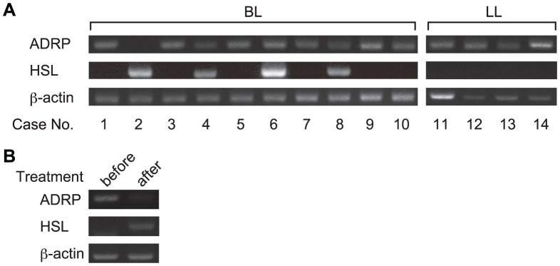
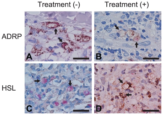
Mycobacterium leprae (M. leprae) lives and replicates within macrophages in a foamy, lipid-laden phagosome. The lipids provide essential nutrition for the mycobacteria, and M. leprae infection modulates expression of important host proteins related to lipid metabolism. Thus, M. leprae infection increases the expression of adipophilin/adipose differentiation-related protein (ADRP) and decreases hormone-sensitive lipase (HSL), facilitating the accumulation and maintenance of lipid-rich environments suitable for the intracellular survival of M. leprae. HSL levels are not detectable in skin smear specimens taken from leprosy patients, but re-appear shortly after multidrug therapy (MDT). This study examined the effect of MDT components on host lipid metabolism in vitro, and the outcome of rifampicin, dapsone and clofazimine treatment on ADRP and HSL expression in THP-1 cells. Clofazimine attenuated the mRNA and protein levels of ADRP in M. leprae-infected cells, while those of HSL were increased. Rifampicin and dapsone did not show any significant effects on ADRP and HSL expression levels. A transient increase of interferon (IFN)-β and IFN-γ mRNA was also observed in cells infected with M. leprae and treated with clofazimine. Lipid droplets accumulated by M. leprae-infection were significantly decreased 48 h after clofazimine treatment. Such effects were not evident in cells without M. leprae infection. In clinical samples, ADRP expression was decreased and HSL expression was increased after treatment. These results suggest that clofazimine modulates lipid metabolism in M. leprae-infected macrophages by modulating the expression of ADRP and HSL. It also induces IFN production in M. leprae-infected cells. The resultant decrease in lipid accumulation, increase in lipolysis, and activation of innate immunity may be some of the key actions of clofazimine.
Conflict of interest statement
The authors have declared that no competing interests exist.
Figures
References
-
- World Health Organization (2011) Leprosy update, 2011. Wkly Epidemiol Rec 86: 389–399. - PubMed
-
- Ridley DS, Jopling WH (1966) Classification of leprosy according to immunity: A five-group system. Int J Lepr Other Mycobact Dis 34: 255–273. - PubMed
-
- Mattos KA, Lara FA, Oliveira VG, Rodrigues LS, D'Avila H, et al. (2011) Modulation of lipid droplets by Mycobacterium leprae in Schwann cells: a putative mechanism for host lipid acquisition and bacterial survival in phagosomes. Cell Microbiol 13: 259–273. - PubMed
-
- Mattos KA, Oliveira VG, D'Avila H, Rodrigues LS, Pinheiro RO, et al. (2011) TLR6-driven lipid droplets in Mycobacterium leprae-infected Schwann cells: immunoinflammatory platforms associated with bacterial persistence. J Immunol 187: 2548–2558. - PubMed
-
- Cardona PJ, Llatjos R, Gordillo S, Diaz J, Ojanguren I, et al. (2000) Evolution of granulomas in lungs of mice infected aerogenically with Mycobacterium tuberculosis . Scand J Immunol 52: 156–163. - PubMed
Publication types
MeSH terms
Substances
LinkOut - more resources
Full Text Sources
Research Materials
