Review
. 2013;18(1-2):43-73.
doi: 10.1615/critrevoncog.v18.i1-2.40.

On the origin of cancer metastasis

Affiliations
Review

On the origin of cancer metastasis

Thomas N Seyfried et al. Crit Rev Oncog. 2013.

Abstract

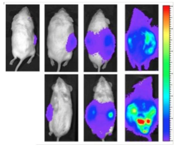
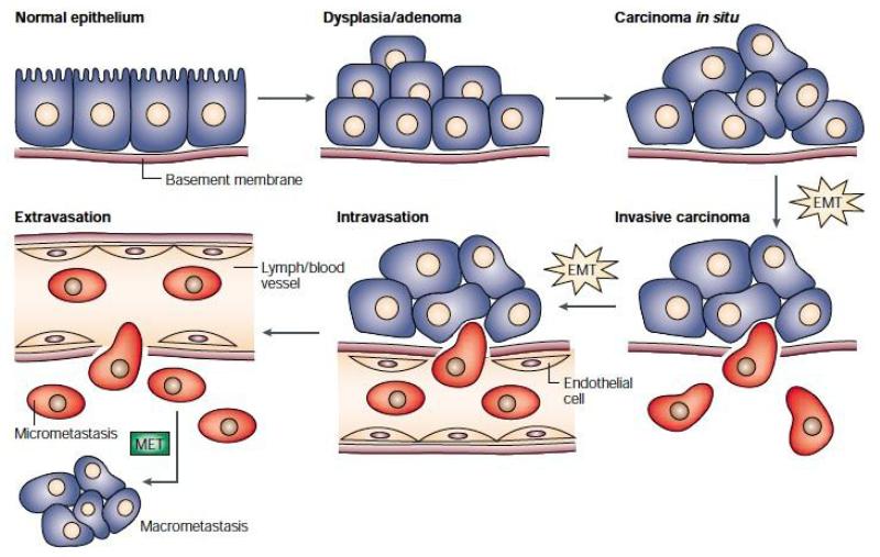
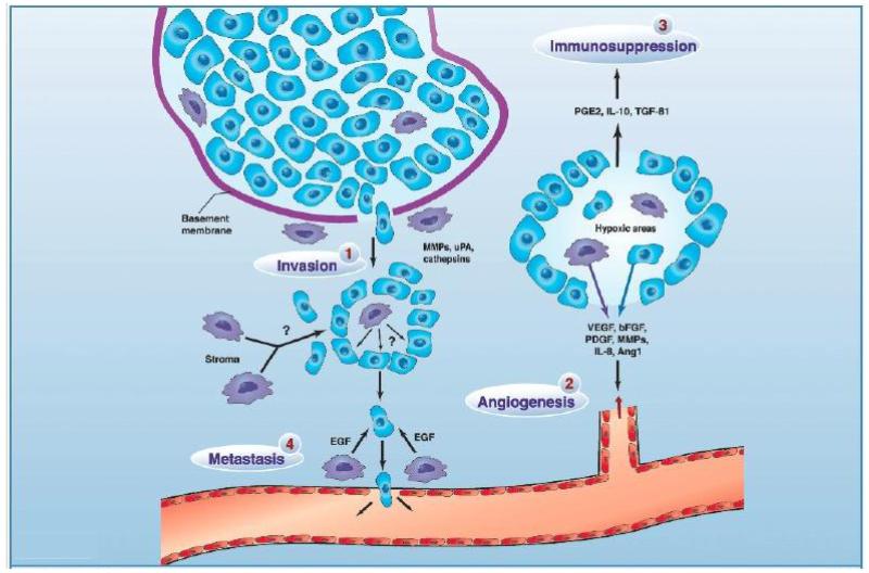
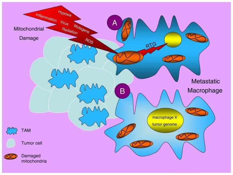
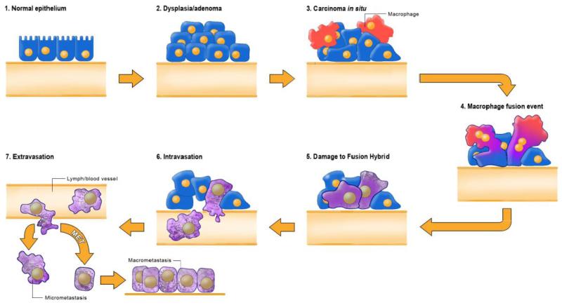
Metastasis involves the spread of cancer cells from the primary tumor to surrounding tissues and to distant organs and is the primary cause of cancer morbidity and mortality. In order to complete the metastatic cascade, cancer cells must detach from the primary tumor, intravasate into the circulatory and lymphatic systems, evade immune attack, extravasate at distant capillary beds, and invade and proliferate in distant organs. Currently, several hypotheses have been advanced to explain the origin of cancer metastasis. These involve an epithelial mesenchymal transition, an accumulation of mutations in stem cells, a macrophage facilitation process, and a macrophage origin involving either transformation or fusion hybridization with neoplastic cells. Many of the properties of metastatic cancer cells are also seen in normal macrophages. A macrophage origin of metastasis can also explain the long-standing "seed and soil" hypothesis and the absence of metastasis in plant cancers. The view of metastasis as a macrophage metabolic disease can provide novel insight for therapeutic management.


Figures

FIGURE 1
FIGURE 1
Systemic metastasis of the VM-M3/Fluc tumor cells grown in the inbred VM mouse. Whole body view of bioluminescence from metastatic VM-M3 tumor cells. VM-M3 tumor cells, containing the firefly luciferase gene, were implanted subcutaneously on the flank of a syngeneic VM mouse on day 0 as we described in (223). Bioluminescent signal from the metastatic cells was measured in live mice using IVIS Lumina system (Caliper LS). Bioluminescence appeared throughout the mouse after 23 days indicative of widespread systemic dissemination of metastatic cells. The mouse is shown in prone position at 3, 10, 17 and 23 days (left to right) after subcutaneous flank implantation of VM-M3/Fluc tumor cells. The bottom row shows the mouse in supine position at those days. Bioluminescent cells were also detected ex vivo in multiple organ systems of the VM mouse host. Source: Reprinted with modification from.
FIGURE 2
FIGURE 2
The epithelial-mesenchymal transition and mesenchymal-epithelial transition (MET) model of tumor metastasis. According to Jean Paul Thiery, normal epithelia lined by a basement membrane can proliferate locally to give rise to an adenoma. Further transformation by epigenetic changes and genetic alterations leads to a carcinoma in situ, still outlined by an intact basement membrane. Further alterations can induce local dissemination of carcinoma cells, possibly through an EMT, as the basement membrane becomes fragmented. The invasive carcinoma cells (red) then intravasate into lymph or blood vessels, allowing their passive transport to distant organs. At secondary sites, solitary carcinoma cells extravasate, remain solitary (micrometastasis), or form a new carcinoma through an MET. Reprinted with permission from.
FIGURE 3
FIGURE 3
The role of different TAM subpopulations in tumor progression. 1, invasion: TAM secrete a variety of proteases to breakdown the basement membrane around areas of proliferating tumor cells (e.g., ductal carcinoma in situ in the breast), thereby prompting their escape into the surrounding stroma where they show deregulated growth. 2, angiogenesis: In areas of transient (avascular) and chronic (perinecrotic) tumor hypoxia, macrophages cooperate with tumor cells to induce a vascular supply for the area by up-regulating a number of angiogenic growth factors and enzymes. These diffuse away from the hypoxic area and, together with other pro-angiogenic stimuli in the tumor microenvironment, stimulate endothelial cells in neighboring, vascularized areas to migrate, proliferate, and differentiate into new vessels. 3, immunosuppression: Macrophages in hypoxic areas secrete factors that suppress the antitumor functions of immune effectors within the tumor. 4, metastasis: A subpopulation of TAM associated with tumor vessels secretes factors like epidermal growth factor (EGF) to guide tumor cells in the stroma toward blood vessels where they then escape into the circulation. TAM secrete growth factors in the stromal compartment to stimulate tumor cell division and/or undefined factors that promote tumor cell motility. Reprinted with permission from reference.
FIGURE 4
FIGURE 4
Proposed mechanisms of macrophage transformation and metastasis. The tumor microenvironment consists of numerous mitochondria damaging elements, which could impair mitochondria energy production in TAM and tissue macrophages. This would eventually produce genetic instability through the mitochondrial stress or retrograde signaling (RTG) response (A)., Fusions between macrophages or between macrophages and cancer stem cells could result in cells expressing both the tumor and macrophage genomes (B). The end result would be cells that can survive in hypoxic environments, can proliferate, and can spread to multiple sites through the circulation. Source: Reprinted with permission from.
FIGURE 5
FIGURE 5
Fusion hybrid hypothesis of cancer cell metastasis. According to our hypothesis, metastatic cancer cells arise following direct transformation or following fusion hybridization between neoplastic epithelial cells and myeloid cells (macrophages). Macrophages are known to invade in situ carcinoma as if it were an unhealed wound. This creates a protracted inflammatory microenvironment leading to fusion hybridization between the neoplastic epithelial cell and the macrophage. Fusion hybridization can explain the phenomenon of EMT without invoking new mutations. Inflammation damages mitochondria leading to enhanced fermentation and acidification of the microenvironment. Mitochondrial damage leading to respiratory insufficiency becomes the driver for the neoplastic transformation of the epithelial cell and of the fusion hybrids (Figure 4). As macrophages are already mesenchymal cells that naturally possess the capability to enter (intravasate) and exit (extravasate) the circulation, the neoplastic fusion hybrid will behave as a rogue macrophage. The fusogenic properties of macrophage cells can also explain how metastatic cells can recapitulate the epithelial characteristics of the primary tumor at secondary micro-metastatic growth sites. This process can explain the phenomenon of MET without invoking a mutation suppression mechanism. Reprinted with permission from Seyfried and Ling.

References

    1. Tarin D. Cell and tissue interactions in carcinogenesis and metastasis and their clinical significance. Sem Cancer Biol. 2011;21(2):72–82. - PubMed
    1. Chambers AF, Groom AC, MacDonald IC. Dissemination and growth of cancer cells in metastatic sites. Nature Rev Cancer. 2002;2(8):563–72. - PubMed
    1. Fidler IJ. The pathogenesis of cancer metastasis: the ‘seed and soil’ hypothesis revisited. Nature Rev Cancer. 2003;3(6):453–8. - PubMed
    1. Welch DR. Defining a cancer metastasis. Amer Assoc Cancer Res. Education Book. 2006:111–5.
    1. Lazebnik Y. What are the hallmarks of cancer? Nature Rev Cancer. 2010;10(4):232–3. - PubMed

Publication types

MeSH terms

LinkOut - more resources