Review
. 2013 Apr;36(4):227-36.
doi: 10.1016/j.tins.2012.11.001. Epub 2012 Dec 11.

Neuronal gap junctions: making and breaking connections during development and injury

Affiliations
Review

Neuronal gap junctions: making and breaking connections during development and injury

Andrei B Belousov et al. Trends Neurosci. 2013 Apr.

Abstract

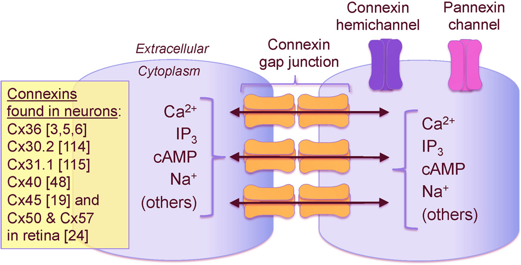
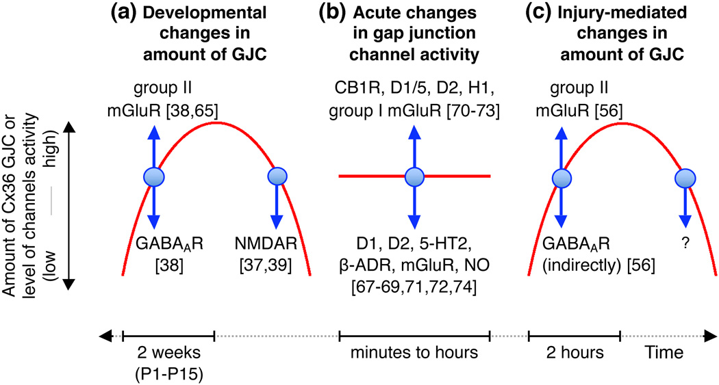
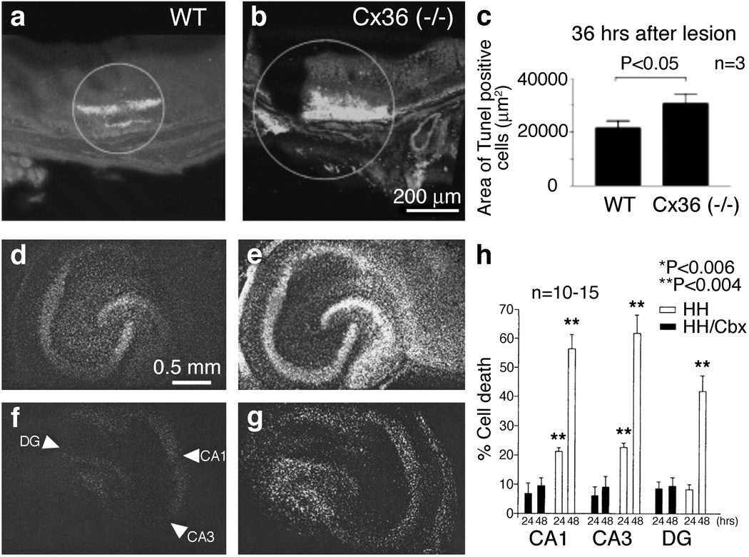
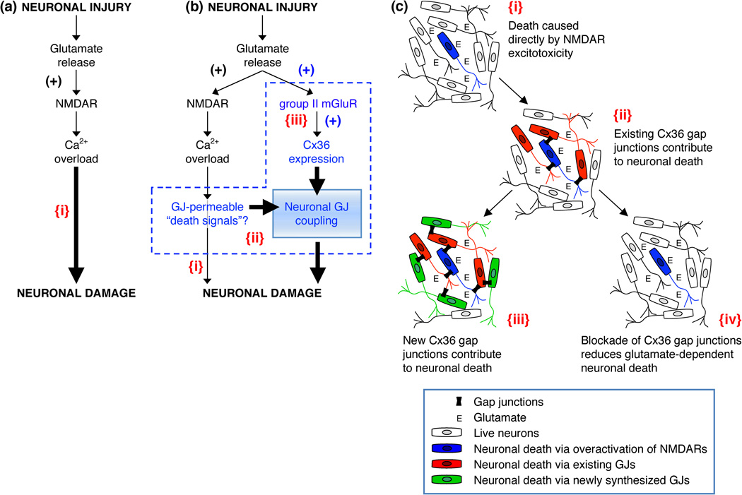
In the mammalian central nervous system (CNS), coupling of neurons by gap junctions (i.e., electrical synapses) and the expression of the neuronal gap junction protein, connexin 36 (Cx36), transiently increase during early postnatal development. The levels of both subsequently decline and remain low in the adult, confined to specific subsets of neurons. However, following neuronal injury [such as ischemia, traumatic brain injury (TBI), and epilepsy], the coupling and expression of Cx36 rise. Here we summarize new findings on the mechanisms of regulation of Cx36-containing gap junctions in the developing and mature CNS and following injury. We also review recent studies suggesting various roles for neuronal gap junctions and in particular their role in glutamate-mediated neuronal death.


Figures

Figure 1
Figure 1. Schematic drawing of connexin gap junctions, connexin hemichannels and pannexin channels
The gap junction channels are made of two hemichannels (one in each apposed membrane) and each hemichannel consists of six connexin subunits. Connexins forming the gap junction channels in neurons are indicated on the figure. Also shown are some of the molecules and ions that pass through gap junctions [1,111]. In addition to gap junctions, unopposed connexin hemichannels and pannexin channels (which, however, do not form gap junctions) also can be expressed by neurons [8,11,12].
Figure 2
Figure 2. Neuronal GJC in the CNS
This figure schematically illustrates conclusions from studies on the role of neurotransmitters and their receptors in (a) developmental, (b) acute, and (c) injury-mediated changes in neuronal GJC. In a and c, red lines represent the increase (upward phase) and decrease (downward phase) in the amount of neuronal GJC and expression of Cx36 during development and neuronal injury; note the differences in duration of developmental (ie. 2 weeks; [–39,65]) and injury-mediated (ie. 2 hours; [56]) increases in GJC. In b, the red line represents background activity of multiple neuronal gap junctions relative to which modulatory changes in the activity occur. In all figures, blue arrows show the direction of change in GJC after activation of the indicated neurotransmitter receptors. Abbreviations: α -ADR, α -adrenoreceptor; CB1R, cannabinoid type 1 receptor; D1, D2 and D1/5, dopamine receptors; GABAAR, GABAA receptor; H1, histamine receptor; 5-HT2, serotonin receptor; mGluR, metabotropic glutamate receptor; NMDAR, NMDA receptor; NO, nitric oxide; P1 and P15, postnatal days 1 and 15.
Figure 3
Figure 3. Is neuronal GJC pro-death or pro-survival?
(a–c) Infrared laser photocoagulation in mouse retina caused retinal trauma, resulting in substantial neuronal death in the retina of wild-type (WT) mice (a) [60]. Such neuronal death was significantly higher in Cx36 knockout (−/−) mice (b). Micrographs of retinas showing TUNEL staining (that detects apoptotic death; a,b) and quantitation (c) are shown. The analysis was done 36 hours after the laser lesioning. Because elimination of Cx36 enhances the level of neuronal death, these data suggest that neuronal GJC is pro-survival [60]. (d–h) Hypoxic-hypoglycemic insult (that models ischemia) induces substantial neuronal death that is seen in organotypic rat hippocampal slices 24 (d) and 48 (e) hours after the insult [110]. Such neuronal death is dramatically reduced by carbenoxolone (a non-specific blocker of GJC) at both 24 (f) and 48 (g) hours post-injury. Images of slices stained with propidium iodide (that detects cell death; d–g) and quantitation of cell death in three main hippocampal regions (CA1, CA3, and dentate gyrus, DG; h) are shown. HH, hypoxia-hypoglycemia; HH/Cbx, hypoxia-hypoglycemia plus carbenoxolone (Cbx, 120 µmol/L) treatment. The three main hippocampal regions are marked by arrowheads in panel f. Together with studies by other groups, demonstrating that pharmacological blockade and knockout of Cx36 GJC dramatically reduce neuronal death in models of ischemia and brain trauma [56,100,107], the data presented in d–h suggest that neuronal GJC is pro-death [110]. Adapted, with permission, from [60] (a–c) and [110] (d–h).
Figure 4
Figure 4. Glutamate-dependent excitotoxicity during neuronal injury
(a) Traditional model of the mechanisms for glutamate-dependent excitotoxicity [104,105]. (b) Alternative model of the mechanisms of glutamate-dependent excitotoxicity [56]. (c) This figure illustrates key points of the alternative model. In all figures: {i}, Neuronal death caused directly by overactivation of NMDARs; {ii}, Existing neuronal gap junctions (GJs) contribute substantially to neuronal death caused by overactivation of NMDARs [100,109]; {iii}, New neuronal gap junctions are induced by activation of group II mGluRs and also contribute to glutamate-dependent neuronal death [56]; {iv}, Pharmacological [100] or genetic [56,100] blockade of neuronal GJC reduces glutamate-dependent neuronal death. In a,b: the sign (+) indicates the increase in receptor activity or expression of Cx36. In b: a possibility is illustrated that some Ca2+-dependent molecules (or Ca2+ itself) serve as gap junction-permeable death signals (b{ii}) [111]; Ca2+ overload also directly induces neuronal death (b{i}), however, this is not the main driver of neuronal death, rather the key determinant for expansion of cell death is neuronal GJC (b{ii,iii}). Adapted, with permission, from [56] (a and b).

References

    1. Eugenin EA, et al. The role of gap junction channels during physiologic and pathologic conditions of the human central nervous system. J Neuroimmune Pharmacol. 2012;7:499–518. - PMC - PubMed
    1. Beyer EC, Berthoud VM. Chapter 1: The family of connexin genes. In: Harris A, Locke D, editors. Connexins: A Guide. Humana Press; 2009. pp. 3–26.
    1. Rash JE, et al. Connexin composition in apposed gap junction hemiplaques revealed by matched double-replica freeze-fracture replica immunogold labeling. J Membr Biol. 2012;245:333–344. - PMC - PubMed
    1. Giaume C, Theis M. Pharmacological and genetic approaches to study connexin-mediated channels in glial cells of the central nervous system. Brain Res Rev. 2010;63:160–176. - PubMed
    1. Belluardo N, et al. Expression of connexin36 in the adult and developing rat brain. Brain Res. 2000;865:121–138. - PubMed

Publication types

MeSH terms