. 2013 Apr 1;319(6):850-9.
doi: 10.1016/j.yexcr.2012.12.002. Epub 2012 Dec 10.

Esophageal epithelial and mesenchymal cross-talk leads to features of epithelial to mesenchymal transition in vitro

Affiliations

Esophageal epithelial and mesenchymal cross-talk leads to features of epithelial to mesenchymal transition in vitro

Amanda B Muir et al. Exp Cell Res. .

Abstract

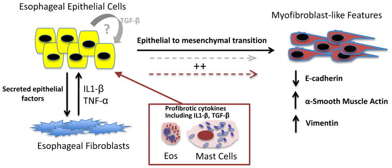
Background: Esophageal fibrosis is a complication of eosinophilic esophagitis (EoE) which has been attributed to both subepithelial fibrosis and to epithelial to mesenchymal transition (EMT), a process by which epithelial cells acquire mesenchymal features. Common to both causes of EoE-fibrosis is the notion that granulocyte-derived TGF-β, induces myofibroblast differentiation of the target cell. To date, the role of esophageal epithelial cells as effector cells in esophageal fibrosis has never been explored. Herein, we investigated consequences of cross-talk between esophageal epithelial cells and fibroblasts, and identified profibrotic cytokines which influence the development of EMT in vitro.

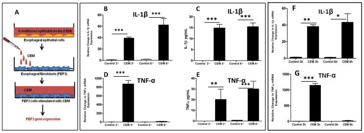
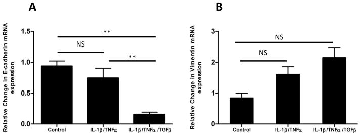
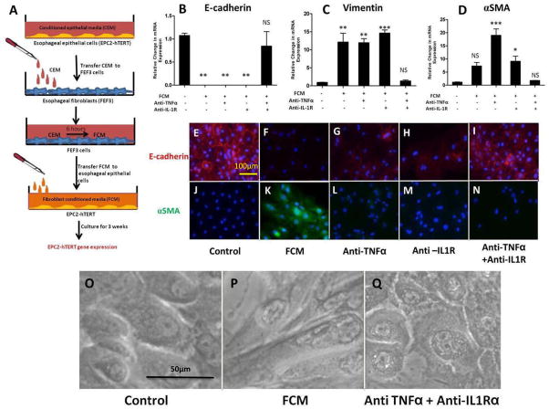
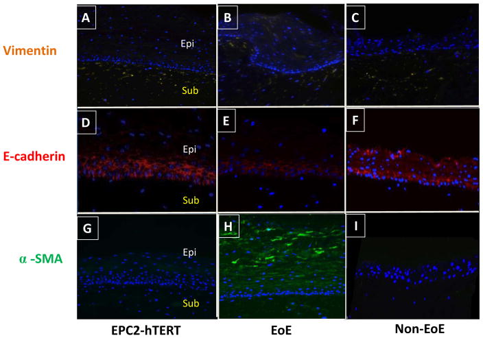
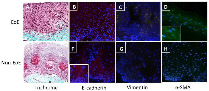
Methods and results: Stimulation of primary fetal esophageal fibroblasts (FEF3) with conditioned media (CEM) from esophageal epithelial cells (EPC2-hTERT), primed FEF3 cells to secrete IL-1β and TNFα, but not TGFβ. To determine whether these cytokines signaled in a paracrine fashion to esophageal epithelial cells, FEF3 cells were stimulated with CEM, followed by transfer of this fibroblast conditioned media (FCM) to EPC2-hTERT cells. Epithelial FCM stimulation increased expression of mesenchymal markers and reduced E-cadherin expression, features of EMT which were TNFα and IL-1β-dependent. Using organotypic culture models, primary EoE epithelial cells exhibited features of EMT compared to non-EoE cells, corresponding to patterns of EMT in native biopsies.

Conclusions: Esophageal epithelial cell and fibroblast cross-talk contributes to esophageal fibrosis. Our results suggest that features of EMT can develop independent of TGF-β and granulocytes, which may have important implications in treatment of EoE.


Conflict of interest statement

Conflicts of Interest: None

Figures

Figure 1
Figure 1. Conditioned epithelial media (CEM) primes fibroblasts to secrete proinflammatory cytokines
A: Schematic of experimental design: Conditioned epithelial media (CEM) from esophageal epithelial cells (EPC2-hTERT) were transferred to fetal esophageal fibroblasts (FEF3 cells). Media was collected at various time points for quantification of FEF3-secreted cytokines, and FEF3 cells were harvested for analysis of gene expression. B, D: mRNA expression of IL-1β and TNFα by FEF3 cells at various time points after CEM stimulation. C, E: Quantification of IL-1β and TNFα secretion by FEF3 cells at various time points following CEM stimulation. F, G: mRNA expression of IL-1β and TNFα expression by primary esophageal fibroblasts (PEF429) from an adolescent EoE patient following stimulation with CEM for various time points. Data shown are representative of at least 3 individual experiments. Error bars represent standard error. * p<0.05, **p<0.01, ***p<0.001, NS = not significant.
Figure 2
Figure 2. Stimulation of esophageal epithelial cell stimulation with IL-1β and TNF-α induces expression of mesenchymal genes and suppresses expression of epithelial markers, features of EMT which are further enhanced by TGF-β
A: mRNA expression of epithelial-specific E-cadherin by EPC2-hTERT cells after 3 weeks of stimulation with combinations of IL-1β, TNFα, and TGF-β. B: Expression of mesenchymal marker vimentin by EPC2-hTERT cells after three weeks of dual cytokine (IL-1β/TNFα) or triple cytokine (IL-1β/TNFα/TGF-β) stimulation. Data shown are representative of at least 3 individual experiments. p-values were calculated based upon comparisons to unstimulated conditions. *p<0.05, **p<0.01, NS = not significant.
Figure 3
Figure 3. Stimulation of esophageal epithelial cells with fibroblast conditioned media (FCM) leads to features of EMT, in an IL-1β and TNFα-dependent fashion
A: Schematic of experimental design: Following stimulation of FEF3 cells with CEM (2 days), this “fibroblast conditioned media” (FCM) was harvested and transferred to fresh EPC2-hTERT cells. After 3 weeks of FCM stimulation in the presence of absence of competitive inhibitors of IL-1β and/or TNFα, EPC2-hTERT cells were harvested for mRNA isolation, or immunolocalization of mesenchymal/epithelial markers. B, C, D: mRNA expression of E-cadherin, vimentin, and αSMA by EPC2-hTERT cells after stimulation with FCM in the presence or absence of anti-TNFα mAb (Remicade) and/or anti-IL-1R (Anakinra). E, J: Constitutive expression of epithelial E-cadherin (red) and αSMA (green) by EPC2-hTERT cells. Nuclei are counterstained with DAPI (blue). F, K: Loss of E-cadherin expression (red) and enhanced αSMA (green) expression by EPC2-hTERT cells following 3 weeks of stimulation with FCM. G, H: Partial recovery of E-cadherin expression by EPC2-hTERT cells stimulated with FCM and anti-TNFα mAb (Remicade) or anti-IL-1R (Anakinra). L, M: Absence of αSMA in EPC2-hTERT cells treated with FCM in the presence anti-TNFα mAb (Remicade) or anti-IL-1R (Anakinra). I, N: Combination of anti-TNFα mAb (Remicade) and anti-IL-1R (Anakinra) protects EPC2-hTERT cells from effects of FCM stimulation. O, P: Morphology of EPC2-hTERT cells before and after FCM stimulation. Q: Effect of anti-TNFα mAb and anti-IL-1R upon EPC2-hTERT morphology. p-values were calculated based upon comparisons to unstimulated conditions. *p<0.05, **p<0.01, ***p<0.001, NS = not significant.
Figure 4
Figure 4. Primary esophageal epithelial cells from an EoE subject exhibit fibrogenic behavior compared to non-EoE control when grown in organotypic cell culture (OTC)
Primary esophageal epithelial cells (passage 3) were harvested from subjects 394 (EoE) and 425 (non-EoE), and seeded onto a matrix of FEF3 cells within a collagen matrix. OTC was also constructed using EPC2-hTERT cells. Following epithelial differentiation and stratification, OTC were harvested for immunolocalization of EMT markers. A, B, C: Expression of mesenchymal marker vimentin (yellow) in EPC2-hTERT, EPC394 (EoE) and EPC425 (non-EoE) OTC. D, E, F: E-cadherin expression (red) in EPC2-hTERT, EPC394 (EoE) and EPC425 (non-EoE) grown in OTC. G, H, I: Expression of α-SMA (green) in OTC constructed using EPC2-hTERT, EPC394, and EPC425 cell lines. In all sections, nuclei are counterstained with DAPI (blue). Epithelial (Epi) and subepithelial (Sub) compartments are labeled. Images shown are at 200X magnification.
Figure 5
Figure 5. Expression of subepithelial collagen and mesenchymal/epithelial markers in native biopsies from EoE and control subjects used for primary esophageal epithelial cell lines
A, E: Trichrome stain of esophageal biopsy from subjects 394 and 425 shows differential subepithelial collagen deposition (blue) in EoE (394) and non EoE (425) subjects. B, F: Reduced epithelial E-cadherin (red) expression in EoE subject compared to normal control. Figure Finset shows magnified detail of E-cadherin in the normal control. C, D: Expression of mesenchymal marker vimentin (yellow) and α-SMA (green) in EoE subject. Figure D inset shows magnified detail of α-SMA expression in the EoE biopsy sample. G, H: Vimentin and α-SMA expression in control subject biopsy. Images are shown at 200X magnification.
Figure 6
Figure 6. Proposed mechanism of esophageal epithelial-mesenchymal cross talk and EMT
Together, our model suggests that in a genetically predisposed host, factors secreted by esophageal epithelial cells prime esophageal fibroblasts to secrete cytokines including IL-1β and TNF-α. Fibroblast-derived cytokines IL-1β and TNF-α, then stimulate adjacent esophageal epithelial cells to lose epithelial markers (E-cadherin) and gain expression of mesenchymal markers including vimentin and α-SMA. Though our in vitro model suggests that while these features of EMT can occur in the absence of TGF-β, granulocyte-derived TGF-β and IL-1β (maroon box and arrows)and potentially autocrine signaling by epithelial-derived TGF-β (gray arrows),may further enhance the development of EMT in vivo.

References

    1. Noel RJ, Putnam PE, Rothenberg ME. Eosinophilic esophagitis. The New England journal of medicine. 2004;351(9):940–1. - PubMed
    1. Scholten D, Reichart D, Paik YH, Lindert J, Bhattacharya J, Glass CK, Brenner DA, Kisseleva T. Migration of fibrocytes in fibrogenic liver injury. Am J Pathol. 2011;179(1):189–98. - PMC - PubMed
    1. Keeley EC, Mehrad B, Strieter RM. The role of fibrocytes in fibrotic diseases of the lungs and heart. Fibrogenesis & tissue repair. 2011;4:2. - PMC - PubMed
    1. Wada T, Sakai N, Sakai Y, Matsushima K, Kaneko S, Furuichi K. Involvement of bone-marrow-derived cells in kidney fibrosis. Clin Exp Nephrol. 2011;15(1):8–13. - PubMed
    1. Hao H, Gabbiani G, Camenzind E, Bacchetta M, Virmani R, Bochaton-Piallat ML. Phenotypic modulation of intima and media smooth muscle cells in fatal cases of coronary artery lesion. Arterioscler Thromb Vase Biol. 2006;26(2):326–32. - PubMed

Publication types

MeSH terms