. 2013 Feb;27(2):224-9.
doi: 10.1038/eye.2012.267. Epub 2012 Dec 14.

Pitfalls in colour photography of choroidal tumours

Affiliations

Pitfalls in colour photography of choroidal tumours

A Schalenbourg et al. Eye (Lond). 2013 Feb.

Abstract

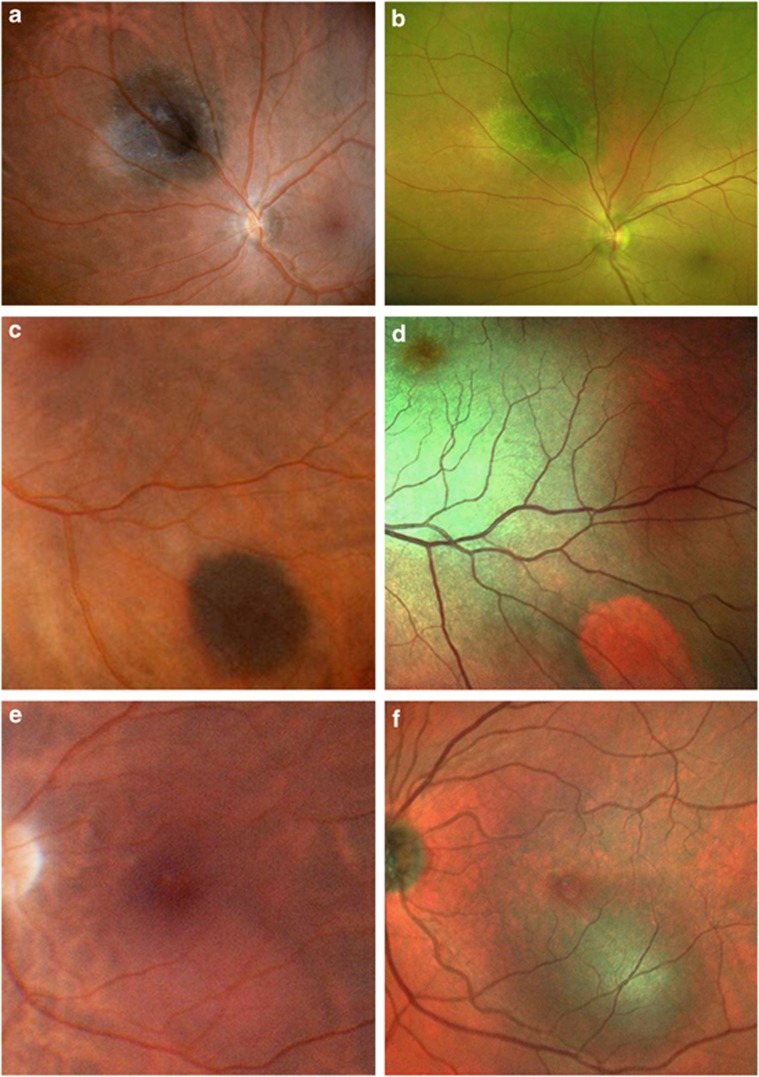
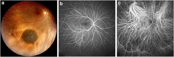
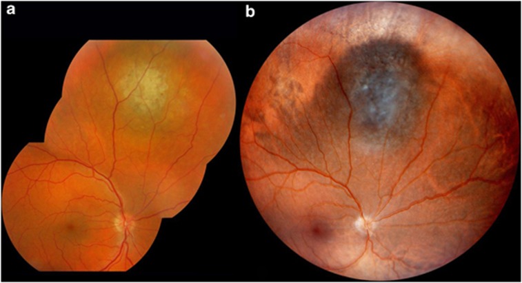
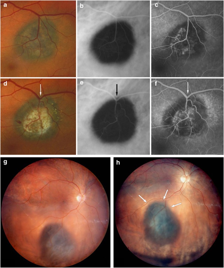
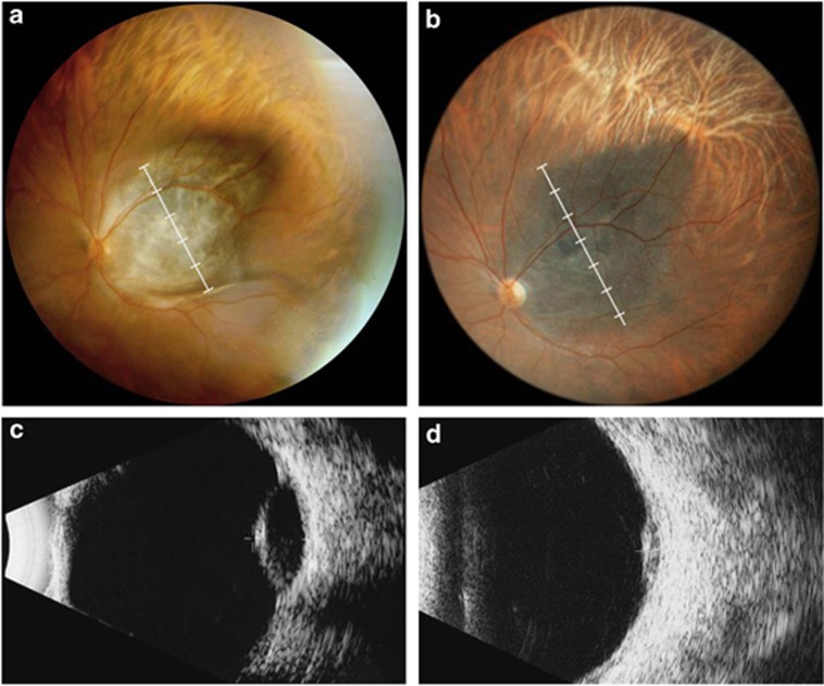
Colour imaging of fundus tumours has been transformed by the development of digital and confocal scanning laser photography. These advances provide numerous benefits, such as panoramic images, increased contrast, non-contact wide-angle imaging, non-mydriatic photography, and simultaneous angiography. False tumour colour representation can, however, cause serious diagnostic errors. Large choroidal tumours can be totally invisible on angiography. Pseudogrowth can occur because of artefacts caused by different methods of fundus illumination, movement of reference blood vessels, and flattening of Bruch's membrane and sclera when tumour regression occurs. Awareness of these pitfalls should prevent the clinician from misdiagnosing tumours and wrongfully concluding that a tumour has grown.


Figures

Figure 1
Figure 1
Different cameras, different colours. Small choroidal melanoma photographed with the Panoret (a) and Optos (b) cameras, the latter producing a false, green colour because it uses only two laser colours. Choroidal naevus imaged with the Panoret (c) and Spectralis Multicolor (d) cameras, the naevus taking an orange-red appearance, suggestive of a haemangioma, with the latter. Circumscribed choroidal haemangioma, inferior to the fovea, imaged using the Panoret (e) and Spectralis Multicolor (f) cameras, the latter misleadingly suggesting a pigmented naevus or melanoma.
Figure 2
Figure 2
The disappearing tumour. A large choroidal naevus, clearly visible on the Panoret picture (a), was examined with fluorescein (b) and ICG (c) angiography (Heidelberg Engineering Spectralis Widefield imaging using a Staurenghi 150° contact lens), on which the melanocytic tumour does not appear at all.
Figure 3
Figure 3
Pseudogrowth evoked by trans-scleral illumination. Colour picture of a large naevus in the superior choroid, taken with transpupillary (a, IMAGEnet Topcon camera) and trans-scleral (b, Panoret camera) illumination on the same day. The tumour appears larger and sharper defined on the latter photo.
Figure 4
Figure 4
Dislocated blood vessels simulating pseudogrowth. Small choroidal naevus complicated by choroidal neovascularisation, treated with laser photocoagulation, which has resulted in traction of the retinal vein towards the tumour, mimicking tumour progression. Standard colour photography (a, d), ICG (b, e), and fluorescein (c, f) angiography, before (a–c) and after (d–f) laser therapy. Panoret fundus picture of a dome-shaped choroidal melanoma with secondary retinal detachment at presentation, with the patient lying down (g) and seated upright (h). Both pictures were taken on the same day. Shifting of the serous retinal detachment has moved the retinal vein closer to the superior tumour border (white arrows).
Figure 5
Figure 5
Regressing tumour, increasing basal diameter. Dome-shaped choroidal melanoma, before (a) and two years after (b) proton therapy (Panoret camera). While the tumour thickness has decreased from 5.8 mm (c) to 2.0 mm (d) on B-scan ultrasonography, the apparent tumour diameter has increased by at least one disc diameter (compare white rulers), because of inward flattening of the underlying sclera.

References

    1. Johnson RN, McDonald HR, Ai E, Jumper JM. Camera artifacts producing the false impression of growth of choroidal melanocytic lesions. Am J Ophthalmol. 2003;135:711–713. - PubMed
    1. Pomerantzeff O. Equator-plus camera. Invest Ophthalmol. 1975;14 (5:401–406. - PubMed
    1. Ducrey N, Pomerantzeff O, Schepens CL, Delori FC, Schneider J. Clinical trials with the Equator-Plus camera. Am J Ophthalmol. 1977;84 (6:840–846. - PubMed
    1. Walsh JB, Garcia JPS, Nieto JC, Rosen RB, Garcia PT, Fradin S.Wide-Angle Digital Fundus Photography: Panoret 1000 vs RetCam 120 Invest Ophthalmol Vis Sci 200243E-Abstract4370
    1. Optomap Product descriptionAvailable at: http://www.optos.com/en-GB/Professionals/Ophthalmology/Product-description/ . Accessed 20 October 2012.