. 2012 Dec 12;32(50):17935-47.
doi: 10.1523/JNEUROSCI.3860-12.2012.

microRNA-21 regulates astrocytic response following spinal cord injury

Affiliations

microRNA-21 regulates astrocytic response following spinal cord injury

Oneil G Bhalala et al. J Neurosci. .

Abstract

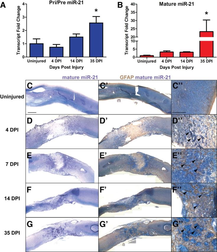
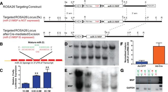
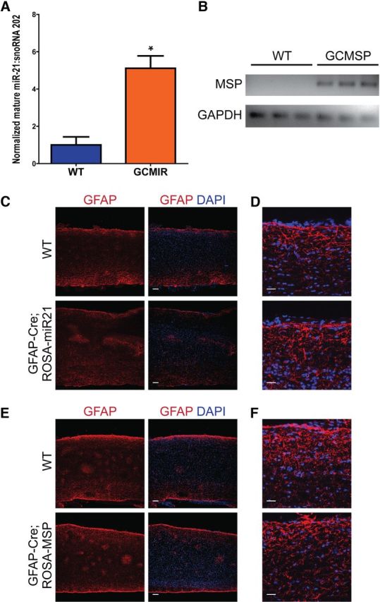
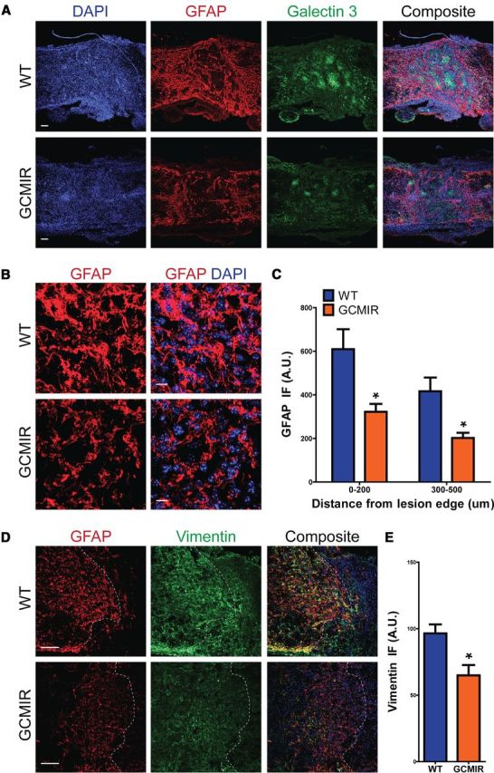
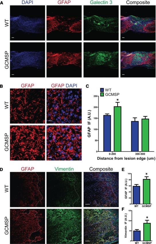
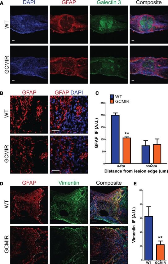
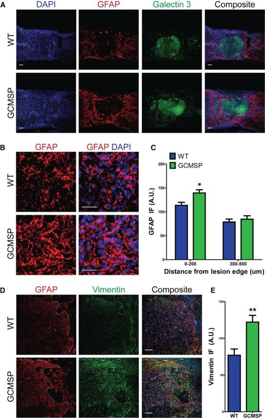
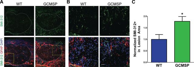
Astrogliosis following spinal cord injury (SCI) involves an early hypertrophic response that serves to repair damaged blood-brain barrier and a subsequent hyperplastic response that results in a dense scar that impedes axon regeneration. The mechanisms regulating these two phases of astrogliosis are beginning to be elucidated. In this study, we found that microRNA-21 (miR-21) increases in a time-dependent manner following SCI in mouse. Astrocytes adjacent to the lesion area express high levels of miR-21 whereas astrocytes in uninjured spinal cord express low levels of miR-21. To study the role of miR-21 in astrocytes after SCI, transgenic mice were generated that conditionally overexpress either the primary miR-21 transcript in astrocytes or a miRNA sponge designed to inhibit miR-21 function. Overexpression of miR-21 in astrocytes attenuated the hypertrophic response to SCI. Conversely, expression of the miR-21 sponge augmented the hypertrophic phenotype, even in chronic stages of SCI recovery when astrocytes have normally become smaller in size with fine processes. Inhibition of miR-21 function in astrocytes also resulted in increased axon density within the lesion site. These findings demonstrate a novel role for miR-21 in regulating astrocytic hypertrophy and glial scar progression after SCI, and suggest miR-21 as a potential therapeutic target for manipulating gliosis and enhancing functional outcome.


Conflict of interest statement

The authors declare no competing financial interests.

Figures

Figure 1.
Figure 1.
miR-21 is expressed after severe spinal cord injury. RNA was extracted from injured spinal cord 4, 14, and 35 DPI, as well as from uninjured spinal cord tissue. A¸ Transcript levels of Pri/Pre miR-21 increase ∼2.5-fold at 35 DPI compared with uninjured tissue. B, Mature miR-21 levels increased by threefold at 4 and 14 DPI compared with uninjured tissue, and by 23-fold at 35 DPI. Error bars indicate SEM. n = 3–4 mice, *p < 0.05 compared with uninjured cord by ANOVA. C–G, in situ hybridization for mature miR-21 revealed expression around lesion area Scale bar, 500 μm. C'–G', Immunohistochemistry for GFAP revealed colocalization with mature miR-21, especially in astrocytes adjacent to lesion site (C”–G” arrowheads). Scale bar, 200 μm.
Figure 2.
Figure 2.
Design and validation of transgenic mice that overexpress or inhibit miR-21. A, Murine primary mir-21 and miR-21 sponge (MSP) sequence was inserted into ROSA26 targeting construct 3′ to PGK-neomycin cassette containing a trimer of the SV40 polyadenylation sequence (3xpA) flanked by loxP sites. The transgene is driven by a CAG promoter with a splice acceptor site (SA). This design allows for controlled expression of primary miR-21 or MSP via recombination of loxP sites by Cre-recombinase protein. (A, AscI; E, EcoRV; P, PacI; R, EcoRI; S, SalI; Sa, SacII; X, XbaI.) See Material and Methods for transgenic mouse generation. B, MSP transcript contains seven repeats of sequence complimentary to mature miR-21. Base pair mismatch minimizes enzymatic degradation of MSP-miR-21 pair. C, Functional inhibition of mature miR-21 by MSP was tested in HEK 293T cells. Increasing amounts of pCMV-d2eGFP-miR21 (M) compared withpCMV-d2eGFP-CXCR4 (C) sequestered miR-21, allowing for increased expression of pCMV-Luc-miR21-B (firefly luciferase). (1C:0M—pCMV-d2eGFP-CXCR4 plasmid only, 0.5C:0.5M—equal amounts of pCMV-d2eGFP-CXCR4 and pCMV-d2eGFP-miR21. 0C:1M—pCMV-d2EGFP-miR21 only.) n = 3, **p < 0.01 by ANOVA, compared with 1C:0M. D, E, Southern blot analyses for 5′ integration of ROSA26 targeting construct for miR-21 and MSP mice, respectively. Five clones and WT are shown for ROSA-miR21. Four clones are shown for MSP. F, ROSA-miR21 ES cells confirmed ROSA26 integration by Southern blot and PCR exhibited ∼8-fold mature miR-21 expression when infected with Ad-Cre compared with Ad-GFP. G, Similarly, confirmed ROSA-MSP ES cells expressed MSP RNA in the presence of Cre (+C) and when RNA was processed by reverse transcriptase (+R). GAPDH served as control for qPCR. Two clones outlined by dashed lines are shown.
Figure 3.
Figure 3.
Modulation of miR-21 does not affect astrocytes under homeostatic conditions. A, Uninjured spinal cords from GCMIR expressed fivefold more mature miR-21compared with WT as detected by qPCR. B, Similarly, MSP expression was detected by qPCR in GCMSP uninjured spinal cords but not WT. C, D, No differences from WT and GCMIR spinal cord astrocytes were observed at low and high magnification, respectively. E, F, Likewise, astrocytes from WT and GCMSP spinal cords were indistinguishable at low and high magnification, respectively. Scale bars: C, E, 100 μm; D, F, 25 μm. *p < 0.02.
Figure 4.
Figure 4.
miR-21 attenuates astrocytic hypertrophy 14DPI after SCI. WT and GCMIR21 mice were analyzed 14 DPI after SCI. A, Longitudinal cross-sections revealed that astrocytes were less hypertrophic and expressed less GFAP (red) in GCMIR21 mice compared with WT. Compaction by astrocytes of activated microglia marker Galectin 3+ (green) was also reduced in GCMIR21 mice compared with WT. Scale bar, 100 μm. B, Magnified images of astrocytes adjacent to lesion showed smaller astrocytes with thinner processes in GCMIR21 mice compared with WT. (GFAP, red; DAPI, blue.) Scale bar, 10 μm. C, Quantification of GFAP immunofluorescence (IF; arbitrary units, A.U.) 0–200 μm and 300–500 μm away from lesion edge. D, Vimentin immunofluorescence (green) is also reduced in astrocytes adjacent to injury site in GCMIR21 mice compared with WT. Dashed white lines indicate lesion edge. Scale bar, 100 μm. E, Quantification of vimentin staining within 200 μm of injury site. n = 4, *p < 0.05 by Student's t test.
Figure 5.
Figure 5.
Inhibition of miR-21 function enhances astrocytic hypertrophy 14DPI after SCI. WT and GCMSP mice were analyzed 14 DPI after SCI. A, In GCMSP injured spinal cord, astrocytes displayed more GFAP (red) immunofluorescence and were hypertrophic with thicker processes compared with WT. Distribution of galectin 3+ microglia (green) was unchanged. Scale bar, 100 μm. B, Higher magnification of astrocytes adjacent to injury demonstrates increased hypertrophy in GCMSP mice. Scale bar, 10 μm. C, Quantification shows increased GFAP staining 0–200 μm from injury site. D, GFAP (red) and vimentin (green) immunofluorescence is increased adjacent to injury site in GCMSP mice. E, F, Quantification of GFAP and vimentin staining, respectively, 0–100 μm from lesion edge. n = 4 mice, *p < 0.05 by Student's t test. Dashed white lines indicate lesion edge.
Figure 6.
Figure 6.
miR-21 reduces GFAP immunofluorescence in injured spinal cords at a chronic stage. WT and GCMIR spinal cords were analyzed 35 DPI after injury. A, Astrocytes expressed less GFAP (red) in GCMIR mice compared with WT around lesion, but still maintained compaction of activated microglia (Galectin 3, green). Scale bar, 100 μm. B, Magnified images (from A) showed equal numbers of astrocytes but reduced GFAP staining (red) in GCMIR. Scale bar, 25 μm. C, GFAP immunofluorescence is reduced with 200 μm of lesion in GCMIR mice. D, GFAP (red) and vimentin (green) is reduced in GCMIR astrocytes adjacent to lesion. E, GCMIR astrocytes showed reduced vimentin immunofluorescence. n = 3–5 mice, **p < 0.01 by Student's t test. Dashed white lines indicate lesion edge.
Figure 7.
Figure 7.
Astrocytic hypertrophy is maintained at chronic stages of injury in GCMSP mice. WT and GCMSP spinal cords were analyzed 35 DPI after injury. A, Astrocytes had more GFAP staining (red) and remained hypertrophic in GCMSP mice, compared with finely processed astrocytes seen in WT cords. Scale bar, 100 μm. B, High magnification shows increased GFAP immunofluorescence (red) and hypertrophy in astrocytes around lesion site in GCMSP cords. Scale bar, 25 μm. C, Quantified GFAP immunofluorescence is increased 0–200 μm from injury edge in GCMSP, but is unchanged 300–500 μm. D, Vimentin immunofluorescence (green) is greatly increased adjacent to lesion in GCMSP, indicating sustained reactive astrocytes. Scale bar, 100 μm. E, Quantified vimentin immunofluorescence is increased in GCMSP injured cords 0–200 μm from lesion edge. n = 3–6 mice, *p < 0.05, **p < 0.01. Dashed white lines indicate lesion edge.
Figure 8.
Figure 8.
Inhibition of miR-21 increases axonal neurofilament expression through the lesion site. A, Staining for the pan-axonal neurofilament marker SMI-312 (green) was increased within the lesion site, demarcated by the astrocytic GFAP border (dashed white line), in GCMSP mice compared with WT mice 35 DPI after injury. Scale bar, 100 μm. B, Higher magnification (yellow boxes demarcated in A) of SMI-312 staining. Scale bar, 25 μm. C, Quantification of percentage of lesion area that is SMI-312+ demonstrated nearly a twofold increase in GCMSP mice normalized to WT. n = 3–6 mice, *p < 0.05 by Student's t test.

References

    1. Ahmed MI, Mardaryev AN, Lewis CJ, Sharov AA, Botchkareva NV. MicroRNA-21 is an important downstream component of BMP signalling in epidermal keratinocytes. J Cell Sci. 2011;124:3399–3404. - PMC - PubMed
    1. Åkerblom M, Sachdeva R, Barde I, Verp S, Gentner B, Trono D, Jakobsson J. MicroRNA-124 is a subventricular zone neuronal fate determinant. J Neurosci. 2012;32:8879–8889. - PMC - PubMed
    1. Barnabé-Heider F, Frisén J. Stem cells for spinal cord repair. Cell stem cell. 2008;3:16–24. - PubMed
    1. Barnabé-Heider F, Göritz C, Sabelström H, Takebayashi H, Pfrieger FW, Meletis K, Frisén J. Origin of new glial cells in intact and injured adult spinal cord. Cell stem cell. 2010;7:470–482. - PubMed
    1. Bartel DP. MicroRNAs: genomics, biogenesis, mechanism, and function. Cell. 2004;116:281–297. - PubMed

Publication types

LinkOut - more resources