. 2012 Dec 12;32(50):17961-9.
doi: 10.1523/JNEUROSCI.3379-12.2012.

Diffusion abnormalities in pediatric mild traumatic brain injury

Affiliations

Diffusion abnormalities in pediatric mild traumatic brain injury

Andrew R Mayer et al. J Neurosci. .

Abstract

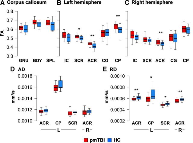
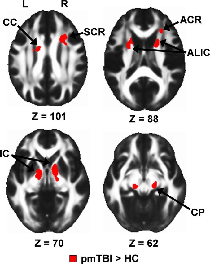
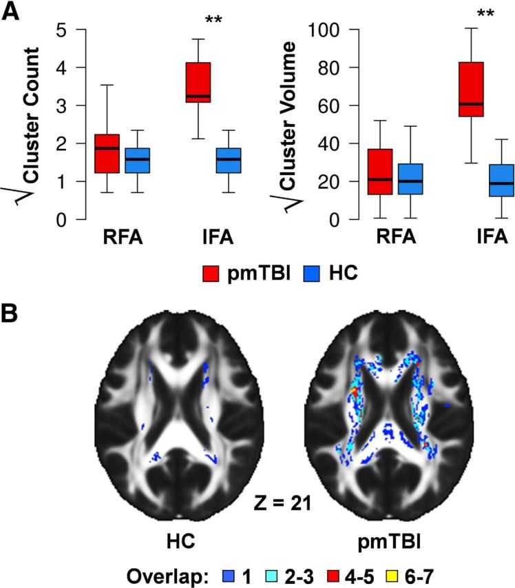
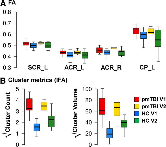
Pediatric mild traumatic brain injury (pmTBI) is the most prevalent neurological insult in children and is associated with both acute and chronic neurobehavioral sequelae. However, little is known about underlying pathophysiology and how injuries change as a function of recovery. Fractional anisotropy, axial diffusivity, and radial diffusivity were examined in 15 semi-acute pmTBI patients and 15 well-matched controls, with a subset of participants returning for a second visit. A novel analytic strategy was applied to capture spatially heterogeneous white matter injuries (lesions) in addition to standard analyses. Evidence of cognitive dysfunction after pmTBI was observed in the domains of attention (p = 0.02, d = -0.92) and processing speed (p = 0.05, d = -0.73) semi-acutely. Region of interest (ROI) and voxelwise analyses indicated increased anisotropic diffusion for pmTBI patients, with an elevated number of clusters with high anisotropy. Metrics of increased anisotropy were able to objectively classify pmTBI from healthy controls at 90% accuracy but were not associated with neuropsychological deficits. Little evidence of recovery in white matter abnormalities was observed over a 4-month interval in returning patients, indicating that physiological recovery may lag behind subjective reports of normality. Increased anisotropic diffusion has been previously linked with cytotoxic edema after TBI, and the magnitude and duration of these abnormalities appear to be greater in pediatric patients. Current findings suggest that developing white matter may be more susceptible to initial mechanical injury forces and that anisotropic diffusion provides an objective biomarker of pmTBI.


Figures

Figure 1.
Figure 1.
This figure presents the FA values from all ROIs for the pmTBI patients (red) and HC (blue) for visit 1. ROIs included the genu (GNU), body (BDY), and splenium (SPL) of the CC, SCR, ACR, the cingulum bundle (CG), CP, and IC (AC). Significant effects are denoted with double asterisks, statistical trends with a single asterisk. D and E present axial diffusivity (AD) and RD values for ROIs that exhibited significant or trend effects, with the y-axis scaled according to millimeters squared per second. Box and whisker plots are used to represent the median and one and a half times the interquartile range.
Figure 2.
Figure 2.
Whole-brain voxelwise results for visit 1 data, presented on an average FA template in Montreal Neurological Institute space (axial slice locations = Z). Selected regions showing increased FA for the pmTBI patients relative to the matched HC included the CC, ACR, SCR, anterior limb of the internal capsule (ALIC), IC, and CP. L, Left; R, right.
Figure 3.
Figure 3.
A presents box plots of cluster metrics that exceed 2 SDs from the HC mean. Specifically, both the number and volume of clusters (minimum of 0.128 ml) that were either two z scores below [reduced FA (RFA)] or two z scores above [increased FA (IFA)] the HC mean were calculated on a voxelwise basis. Each measure was square root transformed to improve normality, and data are presented for both pmTBI patients (red) and HC (blue). Significant effects are denoted with double asterisks. Box and whisker plots are used to represent the median and one and a half times the interquartile range. B displays the distribution of lesions with increased FA summed across participants from both the HC and pmTBI samples selected from a representative slice in Talairach space (Z = 21).
Figure 4.
Figure 4.
A depicts mean FA for returning pmTBI patients (red) and HC (blue) at both visit 1 and visit 2 (pmTBI, yellow; HC, green). Values from ROIs that exhibited either significant or trend differences during visit 1 were examined for longitudinal changes, including the left and right ACR (ACR_L, ACR_R), left CP (CP_L), and left SCR (SCR_L). B displays the longitudinal data for cluster metrics showing increased FA (IFA; for detailed explanation, see Fig. 3) using identical color coding. Box and whisker plots are used to represent the median and one and a half times the interquartile range. For all comparisons, the main effect of group was either significant or at trend levels [pmTBI (red + yellow) > HC (blue + green)] in conjunction with no significant interactions.

References

    1. Adelson PD, Kochanek PM. Head injury in children. J Child Neurol. 1998;13:2–15. - PubMed
    1. Akpinar E, Koroglu M, Ptak T. Diffusion tensor MR imaging in pediatric head trauma. J Comput Assist Tomogr. 2007;31:657–661. - PubMed
    1. Anderson V, Catroppa C, Morse S, Haritou F, Rosenfeld J. Outcome from mild head injury in young children: a prospective study. J Clin Exp Neuropsychol. 2001;23:705–717. - PubMed
    1. Arfanakis K, Haughton VM, Carew JD, Rogers BP, Dempsey RJ, Meyerand ME. Diffusion tensor MR imaging in diffuse axonal injury. Am J Neuroradiol. 2002;23:794–802. - PMC - PubMed
    1. Babikian T, Tong KA, Galloway NR, Freier-Randall MC, Obenaus A, Ashwal S. Diffusion-weighted imaging predicts cognition in pediatric brain injury. Pediatr Neurol. 2009;41:406–412. - PubMed

Publication types

LinkOut - more resources