. 2012 Dec 12;32(50):17977-87.
doi: 10.1523/JNEUROSCI.3069-12.2012.

Dissecting the determinants of light sensitivity in amphioxus microvillar photoreceptors: possible evolutionary implications for melanopsin signaling

Affiliations

Dissecting the determinants of light sensitivity in amphioxus microvillar photoreceptors: possible evolutionary implications for melanopsin signaling

Camilo Ferrer et al. J Neurosci. .

Abstract

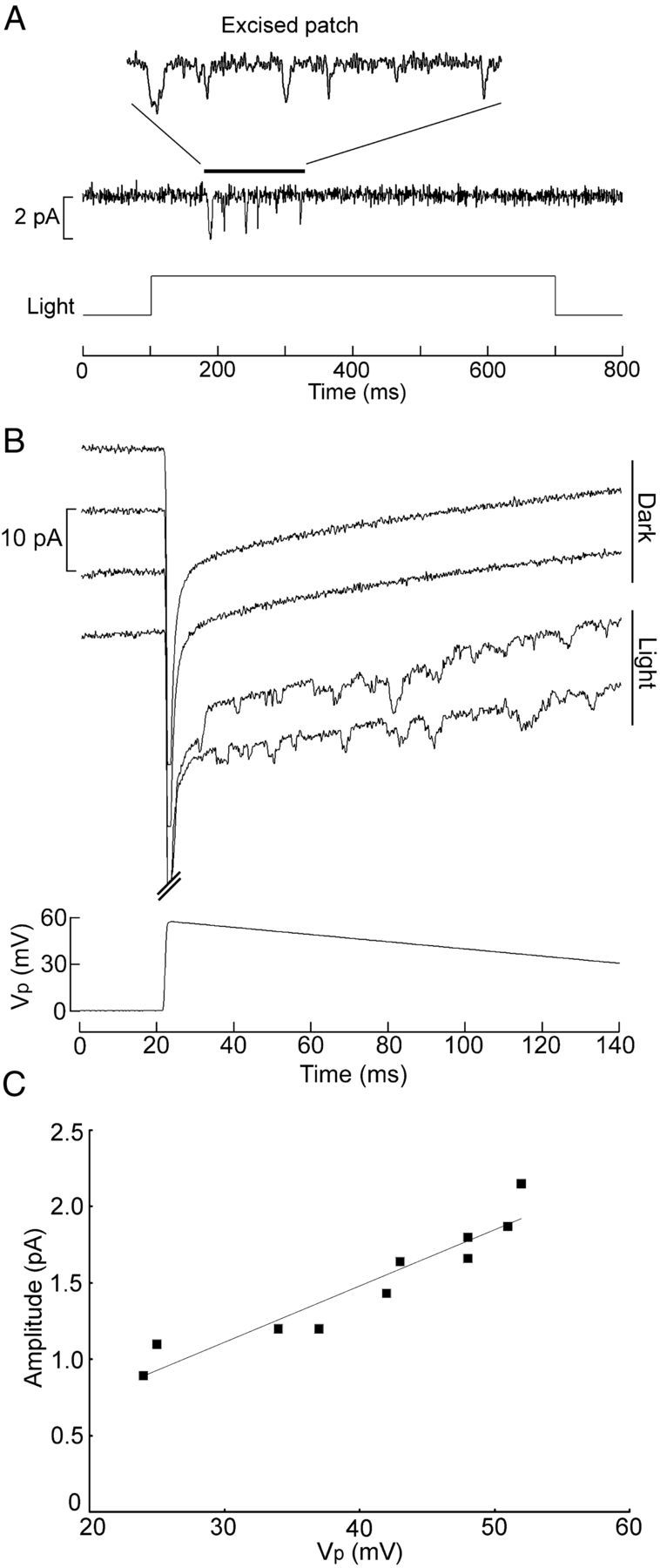
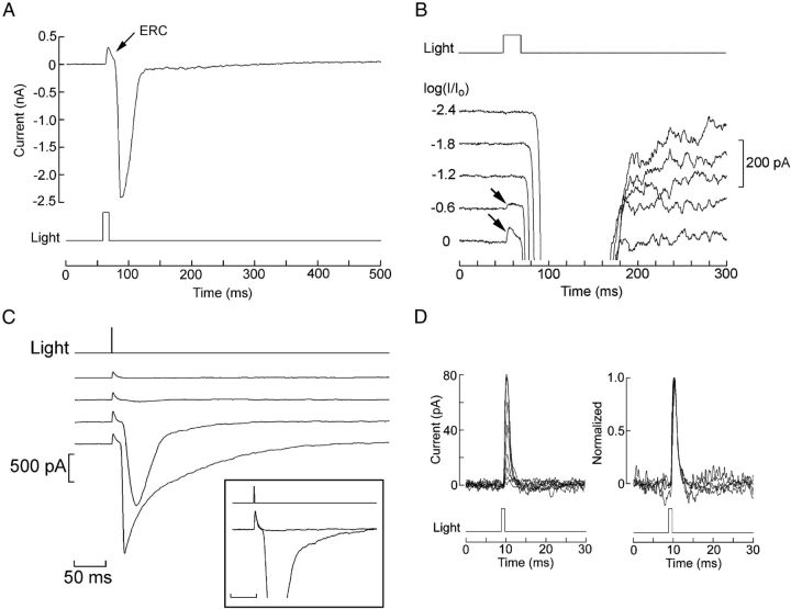
Melanopsin, a photopigment related to the rhodopsin of microvillar photoreceptors of invertebrates, evolved in vertebrates to subserve nonvisual light-sensing functions, such as the pupillary reflex and entrainment of circadian rhythms. However, vertebrate circadian receptors display no hint of a microvillar specialization and show an extremely low light sensitivity and sluggish kinetics. Recently in amphioxus, the most basal chordate, melanopsin-expressing photoreceptors were characterized; these cells share salient properties with both rhabdomeric photoreceptors of invertebrates and circadian receptors of vertebrates. We used electrophysiology to dissect the gain of the light-transduction process in amphioxus and examine key features that help outline the evolutionary transition toward a sensor optimized to report mean ambient illumination rather than mediating spatial vision. By comparing the size of current fluctuations attributable to single photon melanopsin isomerizations with the size of single-channels activated by light, we concluded that the gain of the transduction cascade is lower than in rhabdomeric receptors. In contrast, the expression level of melanopsin (gauged by measuring charge displacements during photo-induced melanopsin isomerization) is comparable with that of canonical visual receptors. A modest amplification in melanopsin-using receptors is therefore apparent in early chordates; the decrease in photopigment expression-and loss of the anatomical correlates-observed in vertebrates subsequently enabled them to attain the low photosensitivity tailored to the role of circadian receptors.


Figures

Figure 1.
Figure 1.
Discrete waves at low illumination intensities. A, Hesse cell voltage clamped at −50 mV was stimulated repetitively with a 400 ms step of light, the intensity of which was increased at 0.3 log increments. Photostimulation elicited inward current waves of approximately constant amplitude and shape, but their frequency increased as a function of light intensity. Unattenuated light intensity 24.6 × 10¹⁵ photons · cm−2 · s−1. B, Wave frequency plotted as a function of photon flux. C, With brighter lights, elementary waves overlap in time, giving rise to larger fluctuation with variable amplitude and multiple peaks (inset). Asterisks mark the stretches that are shown on expanded scale in the inset.
Figure 2.
Figure 2.
Size and speed of the light-evoked elementary waves. A, Stimulation of a Joseph cell with a repetitive flash of light of low intensity (16.7 × 10⁹ photons/cm−2). Distinct quantum bumps (marked with the asterisks) could be identified and their amplitude measured. At this light intensity, a significant fraction of the trials produced no response (data not shown). B, Examples of elementary waves recorded in a Joseph cell. The dashed horizontal lines mark the average baseline current, whereas the cross and the dotted lines indicate the peak and the half-maximal amplitude, respectively. The vertical solid lines delimit the full-width at half-maximal duration.
Figure 3.
Figure 3.
Cell-attached recording of light-activated unitary currents. A, Patch current evoked by a 100 ms flash (13.3 × 10¹⁴ photons/cm−2) in a Joseph cell; the pipette was filled with ASW, and its Vp was set at +80 mV. Shortly after the outward spike, reflecting the depolarization induced by the light, a burst of inward channel currents is observed. B, In the dark, application of depolarization to the patch over a wide range of voltages failed to activate channels. C, The speed of the rising phase of the receptor potential warrants the interpretation that the initial transient observed in on-cell recordings reflects the macroscopic light response picked up via capacitative coupling through the patch: the bottom trace shows membrane potential recorded intracellularly (current-clamp mode) in a Joseph cell; a saturating 1 ms flash (6.3 × 10¹³ photons/cm−2) evoked a rapid transient depolarization. The top trace shows the time derivative of Vm, highlighting the fact that, during the rising phase of the receptor potential, dV/dt reaches 65 V/s; the falling kinetics is significantly slower (min dV/dt = 0.41 V/s).
Figure 4.
Figure 4.
Variability in light-dependent channel density and use of late openings to gauge unitary conductance. A, A patch in a Hesse photoreceptor producing a few isolated channel openings, although the cell was clearly responsive to light, as indicated by the presence of the capacitative transient; the attenuation factor is indicated near each trace (Vp = +50 mV). Unattenuated light intensity 24.6 × 10¹⁵ photons · cm−2 · s−1. B, Voltage changes applied in the dark, spanning a range of 150 mV, failed to produce channel openings. C, Massive, multichannel activity in a Joseph cell, evoked by stimulating light, the intensity of which was progressively increased, as indicated (Vp = +80 mV). The brightest flashes produced responses with a shape reminiscent of the macroscopic photocurrent of the cell. Unattenuated light intensity 11.4 × 10¹⁵ photons · cm−2 · s−1. D, Light-sensitive patch in which channel openings are entirely confined to a late interval after the clearly biphasic IC, that is, after repolarization of the receptor potential (light, 6.2 × 10¹² photons · cm−2 · s−1). E, Activity in a patch containing numerous light-dependent channels. Inset, Expansion of the recording interval 400–500 ms after light onset reveals many clearly resolvable single-channel transitions. F, G, Late channel openings recorded at different pipette potentials in two patches (light, 5.8 × 10¹³ and 6.1 × 10¹⁵ photons · cm−2 · s−1, respectively). H, I–V relationship for the events in G.
Figure 5.
Figure 5.
Excised patches retain phototransduction capabilities. A, Channel activity induced by a step of light in a patch that was excised, and the inner face was perfused with intracellular solution. Vp was set at +50 mV. Notice the absence of the capacitative spike, as one would anticipate from the fact that the trans-patch voltage is under clamp. B, Voltage ramps administered to an excised, perfused patch in either the dark or the presence of a light that was turned on 20 ms before the onset of the ramp. C, I–V relationship for the single-channel currents of the traces in B. The regression line has a slope corresponding to 37 pS. Light intensity, 24.6 × 10¹⁵ photons · cm−2 · s−1.
Figure 6.
Figure 6.
Melanopsin photoisomerization currents. A, Joseph cell stimulated with an intense flash (4.42 × 10¹⁷ photons · cm−2 · s−1) in control conditions (Vh = −50 mV, ASW). A distinct, rapid outward transient precedes the onset of the photocurrent. B, The amplitude of the early transient is graded with light within a range of intensities that saturates the photocurrent. Unattenuated light intensity, 17.6 × 10¹⁷ photons · cm−2 · s−1. C, Differential adaptation of the photocurrent. Repetition of an intense 1 ms stimulus (15.9 × 10¹⁴ photons/cm−2) at 1 min intervals progressively desensitizes the photocurrent, whereas the early transient survives. Inset, Traces 1 and 3 superimposed and normalized. D, The time course of the early transient is independent of stimulus intensity. Left, Superimposition of eight current traces recorded in a Joseph cell stimulated with 1 ms flashes of increasing intensity (0.5–8 × 10¹⁴ photons/cm−2, at ∼2× increments). Right, Normalization shows the invariance of their shape. The two smallest responses were omitted to avoid excessive noise.
Figure 7.
Figure 7.
Functional quantification of melanopsin expression. A, Separation of the ERC from the photocurrent by equimolar substitution of extracellular Na with NMDG and depolarization of the membrane to −30 mV. The late photocurrent is virtually eliminated, whereas the rapid outward component survives. B, Average charge displacement as a function of light intensity, obtained by integration of the ERC. Open symbols, Joseph cells (n = 5–9 per point). Filled symbols, Hesse cells (n = 4–5 per point). Error bars indicate SEM. C, Comparison of the saturating ERC in a Joseph cell, a rhabdomeric photoreceptor from Lima scabra, and a ciliary photoreceptor from Pecten irradians. D, Average charge displacement in the two amphioxus microvillar photoreceptor types (Hesse, n = 9; Joseph, n = 20) and in visual cells isolated from the retina of Lima (n = 11) and Pecten (n = 6).

References

    1. Angueyra JM, Pulido C, Malagón G, Nasi E, Gomez MdP. Melanopsin-expressing amphioxus hotoreceptors transduce light via a phospholipase C signaling cascade. PLoS One. 2012;7:e29813. - PMC - PubMed
    1. Arendt D. Evolution of eyes and photoreceptor cell types. Int J Dev Biol. 2003;47:563–571. - PubMed
    1. Arendt D, Tessmar K, de Campos-Baptista MI, Dorresteijn A, Wittbrodt J. Development of pigment-cup eyes in the polychaete Platynereis dumerilii and evolutionary conservation of larval eyes in Bilateria. Development. 2002;129:1143–1154. - PubMed
    1. Bacigalupo J, Lisman JE. Single channel currents activated by light in Limulus ventral photoreceptors. Nature. 1983;304:268–270. - PubMed
    1. Baylor DA, Lamb TD, Yau KW. Responses of retinal rods to single photons. J Physiol. 1979;288:613–634. - PMC - PubMed

Publication types

LinkOut - more resources