. 2013 Feb 15;126(Pt 4):1009-20.
doi: 10.1242/jcs.119826. Epub 2012 Dec 13.

CXCL12-CXCR4 signaling is required for the maintenance of mouse spermatogonial stem cells

Affiliations

CXCL12-CXCR4 signaling is required for the maintenance of mouse spermatogonial stem cells

Qi-En Yang et al. J Cell Sci. .

Abstract

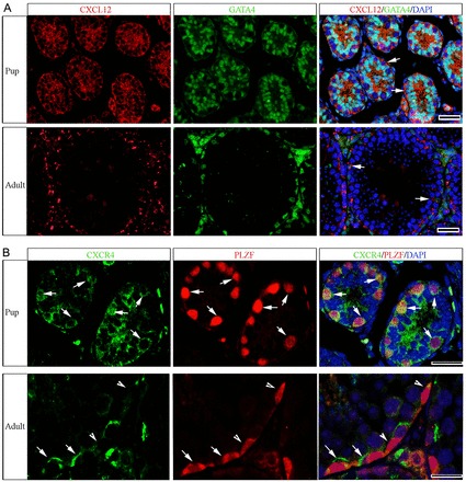
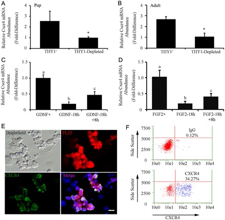
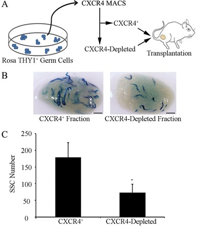
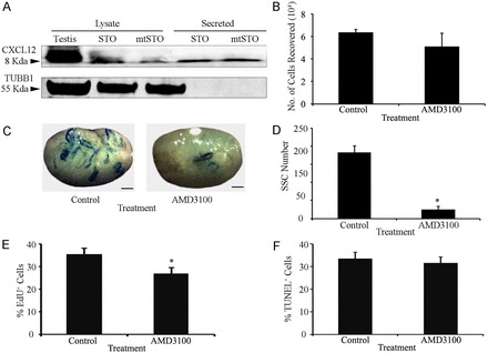
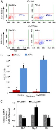
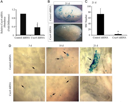
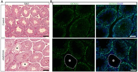
Continual spermatogenesis relies on the activities of a tissue-specific stem cell population referred to as spermatogonial stem cells (SSCs). Fate decisions of stem cells are influenced by their niche environments, a major component of which is soluble factors secreted by support cells. At present, the factors that constitute the SSC niche are undefined. We explored the role of chemokine (C-X-C motif) ligand 12 (CXCL12) signaling via its receptor C-X-C chemokine receptor type 4 (CXCR4) in regulation of mouse SSC fate decisions. Immunofluorescent staining for CXCL12 protein in cross sections of testes from both pup and adult mice revealed its localization at the basement membrane of seminiferous tubules. Within the undifferentiated spermatogonial population of mouse testes, a fraction of cells were found to express CXCR4 and possess stem cell capacity. Inhibition of CXCR4 signaling in primary cultures of mouse undifferentiated spermatogonia resulted in SSC loss, in part by reducing proliferation and increasing the transition to a progenitor state primed for differentiation upon stimulation by retinoic acid. In addition, CXCL12-CXCR4 signaling in mouse SSCs was found to be important for colonization of recipient testes following transplantation, possibly by influencing homing to establish stem-cell niches. Furthermore, inhibition of CXCR4 signaling in testes of adult mice impaired SSC maintenance, leading to loss of the germline. Collectively, these findings indicate that CXCL12 is an important component of the growth factor milieu of stem cells in mammalian testes and that it signals via the CXCR4 to regulate maintenance of the SSC pool.


Figures

Fig. 1.
Fig. 1.
Expression of CXCL12 and CXCR4 within the seminiferous epithelium of testes from postnatal mice. (A) Representative images of immunofluorescent staining for expression of CXCL12 (red) in cross sections of testes from pup (postnatal day 6) and adult (2 months old) mice. Staining is associated with Sertoli cells (arrows) that are labeled by staining for the marker GATA4 (green) and appeared to be concentrated at the basement membrane of seminiferous tubules. (B) Representative images of immunofluorescent staining for expression of CXCR4 (green) in cross sections of testes from pup and adult mice. Staining is observed in cells along the basement membrane that co-stain (red) for expression of the undifferentiated spermatogonial marker PLZF (arrows). Germ cells that stain for PLZF expression but not CXCR4 can also be observed (arrowheads). DAPI was used to stain DNA. Scale bars: 20 µm for all images.
Fig. 2.
Fig. 2.
Expression of Cxcr4 mRNA in the undifferentiated spermatogonial population of mouse testes and regulation by the growth factors influencing SSC self-renewal. (A,B) qRT-PCR analysis for relative Cxcr4 transcript abundance in freshly isolated THY1-positive (THY1+) and THY1-depleted cell fractions from pups (postnatal day 6) and adult (2-month old) mice. Data are means ± s.e.m.; *significant difference at P<0.05. (C) qRT-PCR analysis of the relative Cxcr4 transcript abundance in primary cultures of undifferentiated spermatogonia continually supplemented with GDNF (GDNF+), 18 hours after withdrawal of GDNF supplementation (GDNF-18h), and 8 hours after replacement of GDNF following 18 hours withdrawal (GDNF-18h+8h). Data are means ± s.e.m.; different letters denote significant difference at P<0.05. (D) qRT-PCR analysis for relative Cxcr4 transcript abundance in primary cultures of undifferentiated spermatogonia continually supplemented with FGF2 (FGF2+), 18 hours after withdrawal of FGF2 supplementation (FGF2-18h), and 8 hours after replacement of FGF2 following 18 hours withdrawal (FGF2-18h+8h). Data are means ± s.e.m.; different letters denote significant difference at P<0.05. (E) Representative images of immunofluorescent staining of cells expressing CXCR4 (green) and PLZF (red) in primary cultures of undifferentiated spermatogonia. Scale bars: 10 µm. (F) Representative scatter plots from flow cytometric analysis of the percentage of CXCR4+ cells in primary cultures of undifferentiated spermatogonia. The percentage of CXCR4+ cells is indicated in each plot.
Fig. 3.
Fig. 3.
SSC capacity of CXCR4+ undifferentiated spermatogonia. (A) Schematic of the experimental strategy using magnetic-activated cell sorting (MACS) to isolate CXCR4+ cells from primary cultures of THY1+ undifferentiated spermatogonia derived from lacZ-expressing Rosa donor mice. The CXCR4+ and corresponding CXCR4-depleted fractions were transplanted into the seminiferous tubules of recipient mice. (B) Representative images of recipient mouse testes 2 months after transplantation of single-cell suspensions of isolated CXCR4+ or CXCR4-depleted fractions. Each blue segment represents a colony of spermatogonia derived from a transplanted SSC. Scale bars: 2 mm. (C) Comparison of SSC numbers in the CXCR4+ and CXCR4-depleted cell fractions. Data are means ± s.e.m.; n = 3 different cultures and 12 recipient testes. *Significant difference at P<0.05. SSC numbers were derived from X-gal-stained colonies of donor-derived spermatogonia in recipient testes, normalized to the number of cells (105) injected.
Fig. 4.
Fig. 4.
Impact of CXCR4 signaling on maintenance of SSCs in primary cultures of undifferentiated spermatogonia. (A) Representative images of western blot analysis for CXCL12 protein in lysates and conditioned medium from STO and mitotically inactivated (mt) STO feeder cells. TUBB1 was used as a loading control. (B) Quantitative comparison of total germ cell numbers in primary cultures of undifferentiated spermatogonia after 7 days of treatment with the CXCR4-specific inhibitor AMD3100 or vehicle (control). (C) Representative images of recipient mouse testes 2 months after transplantation with lacZ-expressing primary cultures of undifferentiated spermatogonia treated with vehicle or AMD3100 for 7 days. Scale bars: 2 mm. (D) Quantitative comparison of SSC numbers in primary cultures of undifferentiated spermatogonia after 7 days of treatment with AMD3100 or vehicle, using functional germ cell transplantation analysis (12 total recipient testes). (E) Quantitative comparison of the percentage of proliferative cells (EdU+) within the CXCR4+ fraction of primary cultures of undifferentiated spermatogonia treated with the AMD3100 or vehicle (control). (F) Quantitative comparison of the percentage of apoptotic (TUNEL+) cells within the CXCR4+ fraction of undifferentiated primary cultures treated with AMD3100 or vehicle (control) for 3 days. All data are means ± s.e.m. for three different cultures. *Significant difference at P<0.05.
Fig. 5.
Fig. 5.
Impact of CXCR4 signaling on the response to retinoic acid of primary cultures of undifferentiated spermatogonia. (A) Representative scatter plots from flow cytometric analysis of the percentage of cells expressing the differentiating spermatogonial marker KIT in primary cultures of undifferentiated spermatogonia 24 hours after treatment with all-trans retinoic acid (AtRA) or DMSO (vehicle control). Cultures were pre-treated with the CXCR4-specific inhibitor AMD3100 or vehicle 24 hours before addition of AtRA or DMSO to the culture media. The percentage KIT+ cells in each plot is provided. (B) Quantitative comparison of the percentage of KIT+ cells 24 hours after DMSO or AtRA treatment in primary cultures of undifferentiated spermatogonia pre-treated with AMD3100 or vehicle (control). Data are means ± s.e.m. for three different cultures and different characters denote significantly different at P<0.001. (C) qRT-PCR analysis of relative transcript abundance of Plzf, Ngn3 and Kit in primary cultures of undifferentiated spermatogonia treated with AMD3100 or vehicle. Data are means ± s.e.m. for three different cultures. *Significant difference at P<0.05.
Fig. 6.
Fig. 6.
Impact of CXCR4 deficiency on SSC homing and expansion after transplantation. (A) qRT-PCR analysis for relative Cxcr4 transcript abundance in primary cultures of undifferentiated spermatogonia 24 hours after treatment with control shRNA or Cxcr4 shRNA lentivirus. Data are means ± s.e.m. for three different cultures. *Significant difference at P<0.05. (B) Representative images of recipient mouse testes 21 days after transplantation of cultured undifferentiated spermatogonia stably transduced with control or Cxcr4 shRNA lentiviral transgenes. (C) Comparison of SSC numbers in primary cultures of undifferentiated spermatogonia stably transduced with control or Cxcr4 shRNA lentiviral transgenes using functional germ cell transplantation analysis. SSCs were counted in X-gal-stained colonies of donor-derived spermatogonia in recipient testes, normalized to the number of cells (105) injected. Data are means ± s.e.m. for three different cultures and nine total recipient testes for each treatment. *Significant difference at P<0.05. (D) Representative images of seminiferous tubules 3, 10 and 21 days after transplantation with cultured undifferentiated spermatogonia stably transduced with control or Cxcr4 shRNA lentiviral transgenes. Arrows indicate lacZ-expressing donor cells.
Fig. 7.
Fig. 7.
Impact of pharmacological inhibition of CXCR4 signaling on maintenance of spermatogenesis in testes of adult mice. (A) Representative images of Hematoxylin and Eosin (H&E) stained cross sections from testes of adult mice 28 days after receiving daily injections of vehicle or the CXCR4-specific inhibitor AMD3100 for 7 days. Spermatogenesis appeared normal in all seminiferous tubules in cross sections of testes from control animals. In contrast, in 8–10% of seminiferous tubules from males treated with AMD3100 (n = 3 different animals) spermatogenesis was disrupted and all germ cell types were absent (asterisks). Scale bars: 50 µm. (B) Representative images of immunofluorescent staining for the germ-cell-specific marker GCNA1 (green) in cross sections of testes from mice treated with the CXCR4 inhibitor AMD3100. Seminiferous tubules completely lacking germ cells with a Sertoli-cell-only phenotype that is indicative of SSC loss are observed (asterisks). DAPI (blue) was used to stain DNA. Scale bars: 50 µm.

References

    1. Ara T., Nakamura Y., Egawa T., Sugiyama T., Abe K., Kishimoto T., Matsui Y., Nagasawa T. (2003). Impaired colonization of the gonads by primordial germ cells in mice lacking a chemokine, stromal cell-derived factor-1 (SDF-1). Proc. Natl. Acad. Sci. USA 100, 5319–5323 10.1073/pnas.0730719100 - DOI - PMC - PubMed
    1. Arai A., Jin A., Yan W., Mizuchi D., Yamamoto K., Nanki T., Miura O. (2005). SDF-1 synergistically enhances IL-3-induced activation of the Raf-1/MEK/Erk signaling pathway through activation of Rac and its effector Pak kinases to promote hematopoiesis and chemotaxis. Cell. Signal. 17, 497–506 10.1016/j.cellsig.2004.09.007 - DOI - PubMed
    1. Bellvé A. R., Cavicchia J. C., Millette C. F., O'Brien D. A., Bhatnagar Y. M., Dym M. (1977). Spermatogenic cells of the prepuberal mouse. Isolation and morphological characterization. J. Cell Biol. 74, 68–85 10.1083/jcb.74.1.68 - DOI - PMC - PubMed
    1. Bowles J., Feng C. W., Spiller C., Davidson T. L., Jackson A., Koopman P. (2010). FGF9 suppresses meiosis and promotes male germ cell fate in mice. Dev. Cell 19, 440–449 10.1016/j.devcel.2010.08.010 - DOI - PubMed
    1. Brinster R. L., Zimmermann J. W. (1994). Spermatogenesis following male germ-cell transplantation. Proc. Natl. Acad. Sci. USA 91, 11298–11302 10.1073/pnas.91.24.11298 - DOI - PMC - PubMed

Publication types