. 2013 Feb 25;25(8):1209-15.
doi: 10.1002/adma.201203824. Epub 2012 Dec 12.

A highly tunable biocompatible and multifunctional biodegradable elastomer

Affiliations

A highly tunable biocompatible and multifunctional biodegradable elastomer

Maria José Nunes Pereira et al. Adv Mater. .
No abstract available


Figures

Figure 1
Figure 1
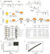
Chemical and mechanical characterization of PGSU elastomers. A) Synthetic scheme for PGSU. B) Synthetic routes for PGSU synthesis under (i) solvent-based and (ii) solvent-free conditions. C) FTIR analysis of PGS pre-polymer and PGSU polymers synthesized under solvent-based and solvent free conditions. D) Summary of mechanical properties and degree of crosslinking for several PGSU derivatives (YM: Young’s modulus, UTS: ultimate tensile strength, El: elongation, n: crosslinking density. E) Typical stress-strain of PGSU-S films and thermally cured PGS elastomer and representative images of PGSU-S 1:0.3 films before and after tensile testing, revealing minimal creep and size/shape changes. F) Stress-strain profile of PGSU-SF 1:0.5 films during 100 cycles of tensile loading shows that the elastomer maintains its tensile properties with minimal creep.
Figure 2
Figure 2
In vivo subcutaneous and cardiac biocompatibility/biodegradation of PGSU elastomers. A) Representative images of H&E and anti-CD68 stained histological sections of the subcutaneous tissue surrounding PGSU-S 1:1 and PLGA polymers (P: polymer implant location, F: fibrous capsule). Scale bar represents 100 μm. B) Characterization of foreign body response to PGSU-S and PLGA implants through qualitative evaluation of the inflammatory infiltrate (from 0 representing no infiltrate, to 4 representing severe infiltrate). C) In vivo degradation profile of PGSU-S films implanted subcutaneously. D) Morphologic evaluation of PGSU-S 1:0.3 and 1:1 cross-sections through SEM (S: polymer surface, CS: polymer cross-section). Scale bar represents 50 μm. E) Representative images of H&E sections of cardiac tissue in contact with PGSU-SF 1:0.3 elastomer following implantation for 1 and 4 weeks (M: myocardium tissue, P: polymer implant location). D) Cardiac function before and 4 weeks after PGSU-SF implantation.
Figure 3
Figure 3
Sustained release of bioactive proteins from PGSU-SF 1:0.3 films. A) Release kinetics (i) and bioactivity (ii) of the lysozyme released from PGSU-SF 1:0.3 porous patches. B) Selective encapsulation of rhodamine and FITC intercalated in PGSU-SF 1:0.3 layers using a spin coating technique. Scale bars represent 100 μm. C) Release kinetics of the model protein BSA sieved to <75 and <32 μm particle size encapsulated within the internal layer of a trilayer spin coated PGSU-SF 1:0.3 film. D) Release kinetics of BSA co-encapsulated with the osmotic agent trehalose (1:1 ratio) and sieved to <32 μm particle size from internal and externals layers of a tri-layer spin coated PGSU-SF 1:0.3 film. Trehalose accelerates protein released by promoting increased water uptake of PGSU-SF 1:0.3 polymers.

References

    1. Serrano MC, Chung EJ, Ameer GA. Adv Funct Mater. 2010;20:192.
    1. Stuckey D, Ishii H, Chen QZ, Boccaccini A, Hansen U, Carr C, Roether J, Jawad H, Tyler D, Ali N, Clarke K, Harding S. Tissue Eng Part B. 2010;16:3395. - PubMed
    1. Wang YD, Ameer GA, Sheppard BJ, Langer R. Nat Biotechnol. 2002;20:602. - PubMed
    1. Guan J, Sacks M, Beckman E, Wagner W. Biomaterials. 2004;25:85. - PubMed
    1. Yang J, Webb AR, Ameer GA. Adv Mater. 2004;16:511.

Publication types

MeSH terms

LinkOut - more resources