. 2013 Jan;137(2):383-96.
doi: 10.1007/s10549-012-2353-5. Epub 2012 Dec 13.

Vimentin DNA methylation predicts survival in breast cancer

Affiliations

Vimentin DNA methylation predicts survival in breast cancer

Jacob Ulirsch et al. Breast Cancer Res Treat. 2013 Jan.

Abstract

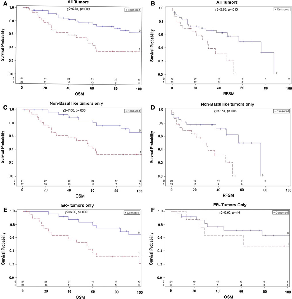
The Vimentin gene plays a pivotal role in epithelial-to-mesenchymal transition and is known to be overexpressed in the prognostically poor basal-like breast cancer subtype. Recent studies have reported Vimentin DNA methylation in association with poor clinical outcomes in other solid tumors, but not in breast cancer. We therefore quantified Vimentin DNA methylation using MALDI-TOF mass spectrometry in breast tumors and matched normal pairs in association with gene expression and survival in a hospital-based study of breast cancer patients. Gene expression data via qRT-PCR in cell lines and oligomicroarray data from breast tissues were correlated with percent methylation in the Vimentin promoter. A threshold of 20 percent average methylation compared with matched normal pairs was set for bivariate and multivariate tests of association between methylation and tumor subtype, tumor histopathology, and survival. Vimentin was differentially methylated in luminal breast cancer cell lines, and in luminal A, luminal B, and HER2-enriched breast tumor subtypes, but was rare in basal-like cell lines and tumors. Increased methylation was strongly correlated with decreased mRNA expression in cell lines, and had a moderate inverse correlation in breast tumors. Vimentin methylation predicted poor overall survival independent of race, subtype, stage, nodal status, or metastatic disease and holds promise as a new prognostic biomarker for breast cancer patients.


Conflict of interest statement

Conflicts of interest The experiments described in this study comply with the current laws of the country in which they were performed. CMP is a major stockholder of Bioclassifier LLC and co-founder and managing partner of University Genomics. The other authors declare no competing interest.

Figures

Fig. 1
Fig. 1
Position of CpG dinucleotides interrogated for methylation. Top Row Base pair positions for the Vimentin amplicon and CG dinucleotides are listed relative to the ATG translational start site (TSS). The TATA box in the Vimentin promoter is at −192 bp. Bottom row Expanded view of the 282-bp amplicon shows each of the 1–17 CG sequences (represented by a circle on top of a vertical line), interrogated for methylation relative to the TSS. The entire sequence was based on human build NCBI 36/hg19, with the Vimentin amplicon spanning bp 17311015–17311296
Fig. 2
Fig. 2
a Cluster analysis of differential Vimentin methylation in breast cancer cell lines. Vimentin DNA methylation was quantified on a mass array platform per CpG unit across the amplicon. Percent methylation is represented on a color continuum from zero percent methylation (gold) to 50 % methylation (black) to 100 % methylation (purple). ER+ and HER2+ luminal cell lines (T47D, MCF7, ZR75 and SKBR3) were hypermethylated while the remaining hormone receptor (HR−) cell lines were hypomethylated. b Vimentin qRT-PCR electrophoresis in breast cancer cell lines. qRT-PCR was carried out in a subset of breast cell lines on an ABI 7500 real-time platform in 20 µl reaction volumes. GAPDH was amplified in tandem and used as the reference control. Vimentin product bands (73 bp) are faint or absent for the luminal, hypermethylated cell lines (lanes 1–4), while robust product bands are seen in the HR− cell lines (lanes 5–11). c Vimentin expression in breast cancer cell lines. qRT-PCR was used to quantify Vimentin expression relative to GAPDH using commercially available ABI TaqMan probes and primers. All breast cell lines were examined in triplicate. Error bars represent the standard error of the mean. HCC1937 was used as the referent cell line, and log10 (RQ) was set at 3.5. Differences between luminal and HR− cell line mRNA expression were highly significant by the student’s t test (t = 10.12; p < 0.00001) and by the Mann–Whitney test (U = 32.0; p < .01)
Fig. 3
Fig. 3
a, b Vimentin methylation in breast tumors and matched tumor normal pairs by molecular subtype. a Breast tumors (n = 81) are listed on the horizontal axis and percent methylation on the vertical axis. Diamonds represent percent methylation for each CpG unit within the Vimentin amplicon. Note Similar percent methylation values for either CpGs 1, 2, 3, 4, 5, 6, 7, 8, 16, and 17 can overlap and may appear as “one diamond.” Tumors are grouped by molecular subtypes assigned from previous oligoarray analysis. (Basal = red, HER2-enriched = pink, Luminal A = Dark Blue, and Luminal B = Light blue). b Top row Methylation of n = 57 tumors and n = 57 matched adjacent normal breast tissue pairs. Differential methylation is seen in tumors, while paired normal tissues have average baseline methylation values of 11 % or less. c ANOVA of Vimentin CpG 17 methylation by molecular subtype. ANOVA was performed in n = 83 tumors + N = 57 matched normal pairs (n = 140 total samples) and grouped by molecular subtype (x axis). The boxplot shows percent methylation distributions where the upper and lower whiskers represent 1.5 times the interquartile range (IQR). Significant differences (p < 0.00001) were observed in percent methylation between tumor subtypes
Fig. 3
Fig. 3
a, b Vimentin methylation in breast tumors and matched tumor normal pairs by molecular subtype. a Breast tumors (n = 81) are listed on the horizontal axis and percent methylation on the vertical axis. Diamonds represent percent methylation for each CpG unit within the Vimentin amplicon. Note Similar percent methylation values for either CpGs 1, 2, 3, 4, 5, 6, 7, 8, 16, and 17 can overlap and may appear as “one diamond.” Tumors are grouped by molecular subtypes assigned from previous oligoarray analysis. (Basal = red, HER2-enriched = pink, Luminal A = Dark Blue, and Luminal B = Light blue). b Top row Methylation of n = 57 tumors and n = 57 matched adjacent normal breast tissue pairs. Differential methylation is seen in tumors, while paired normal tissues have average baseline methylation values of 11 % or less. c ANOVA of Vimentin CpG 17 methylation by molecular subtype. ANOVA was performed in n = 83 tumors + N = 57 matched normal pairs (n = 140 total samples) and grouped by molecular subtype (x axis). The boxplot shows percent methylation distributions where the upper and lower whiskers represent 1.5 times the interquartile range (IQR). Significant differences (p < 0.00001) were observed in percent methylation between tumor subtypes
Fig. 4
Fig. 4
a–f Overall and recurrence free survival by Vimentin methylation status. Kaplan–Meier overall (OS) and recurrence free (RFS) survival analyses were carried out by stratifying breast cancer patients with average Vimentin methylation thresholds above 20 percent (1 = red line) and below 20 percent (0 = blue line) based on relative methylation in matched normal tissues. OS and RFS were recorded in months (horizontal axis). The number of tumors in each analysis varied according to complete data available for the variables under study: a OS in N = 79 breast tumors, b RFS in N = 65 breast tumors, c OS in N = 58 tumors after removing the basal-like tumors from the analysis, d RFS in N = 51 tumors after removing 22 basal-like tumors from the analysis, e OS in N = 47 ER+ tumors only, f OS in N = 32 ER− tumors only. Results for Fig. 4a–e were highly significant and showed breast cancer patients with <20 % average Vimentin methylation survive much longer than those with >20 % methylation (range log rank χ2 = 5.93–7.51, p = 0.015–0.006). Survival differences were not significant in ER− breast tumors (f)

References

    1. Challa AA, Stefanovic B. A novel role of vimentin filaments: binding and stabilization of collagen mRNAs. Mol Cell Biol. 2011;31:3773–3789. - PMC - PubMed
    1. Chen WD, Han ZJ, Skoletsky J, Olson J, Sah J, Myeroff L, Platzer P, Lu S, Dawson D, Willis J, Pretlow TP, Lutterbaugh J, Kasturi L, Willson JK, Rao JS, Shuber A, Markowitz SD. Detection in fecal DNA of colon cancer-specific methylation of the nonexpressed vimentin gene. J Natl Cancer Inst. 2005;97:1124–1132. - PubMed
    1. Coolen MW, Statham AL, Gardiner-Garden M, Clark SJ. Genomic profiling of CpG methylation and allelic specificity using quantitative high-throughput mass spectrometry: critical evaluation and improvements. Nucleic Acids Res. 2007;35:e119. - PMC - PubMed
    1. Costa VL, Henrique R, Danielsen SA, Duarte-Pereira S, Eknaes M, Skotheim RI, Rodrigues A, Magalhaes JS, Oliveira J, Lothe RA, Teixeira MR, Jeronimo C, Lind GE. Three epigenetic biomarkers, GDF15, TMEFF2, and, VIM, accurately predict bladder cancer from DNA-based analyses of urine samples. Clin Cancer Res. 2010;16:5842–5851. - PubMed
    1. Dumont N, Wilson MB, Crawford YG, Reynolds PA, Sigaroudinia M, Tlsty TD. Sustained induction of epithelial to mesenchymal transition activates DNA methylation of genes silenced in basal-like breast cancers. Proc Natl Acad Sci USA. 2008;105:14867–14872. - PMC - PubMed

Publication types