. 2013 Mar;48(3):364-73.
doi: 10.1165/rcmb.2012-0146OC. Epub 2012 Dec 13.

Ciliated cells of pseudostratified airway epithelium do not become mucous cells after ovalbumin challenge

Affiliations

Ciliated cells of pseudostratified airway epithelium do not become mucous cells after ovalbumin challenge

Ana Pardo-Saganta et al. Am J Respir Cell Mol Biol. 2013 Mar.

Abstract

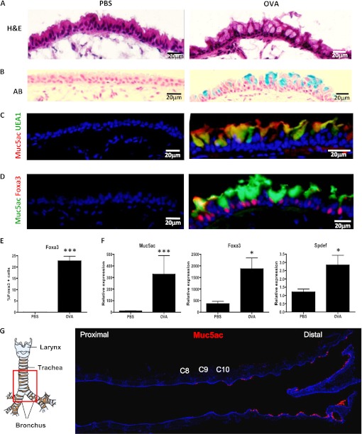
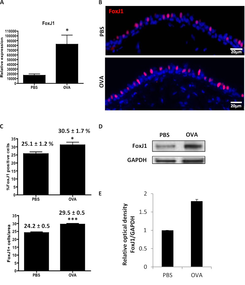
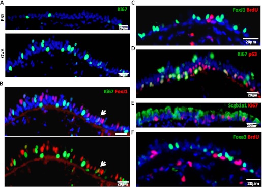
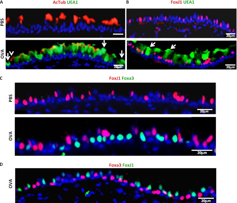
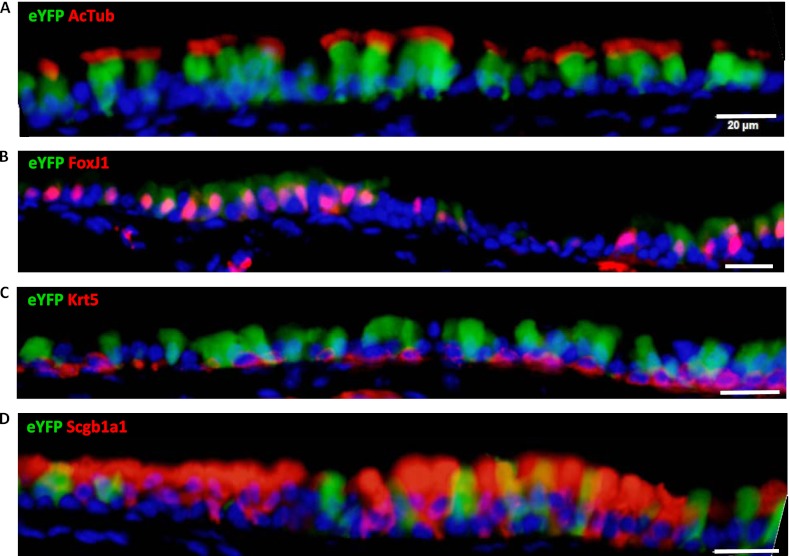
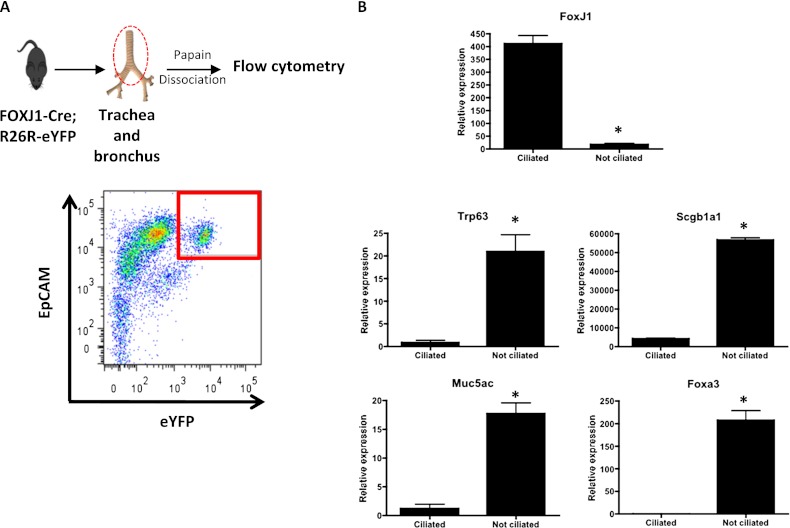
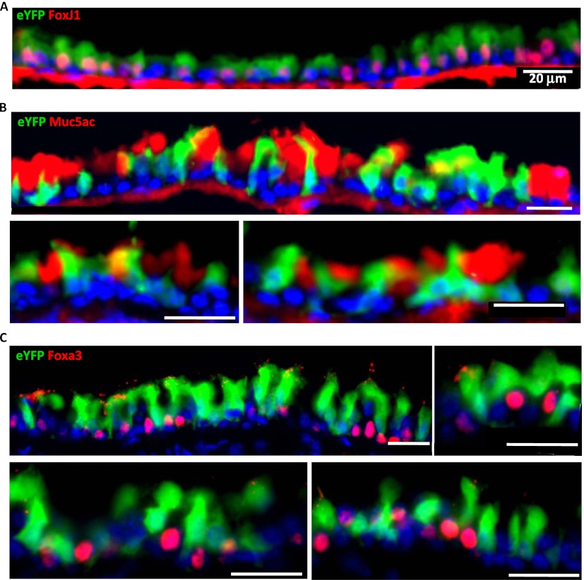
Mucous cell metaplasia is a hallmark of airway diseases, such as asthma and chronic obstructive pulmonary disease. The majority of human airway epithelium is pseudostratified, but the cell of origin of mucous cells has not been definitively established in this type of airway epithelium. There is evidence that ciliated, club cell (Clara), and basal cells can all give rise to mucus-producing cells in different contexts. Because pseudostratified airway epithelium contains distinct progenitor cells from simple columnar airway epithelium, the lineage relationships of progenitor cells to mucous cells may be different in these two epithelial types. We therefore performed lineage tracing of the ciliated cells of the murine basal cell-containing airway epithelium in conjunction with the ovalbumin (OVA)-induced murine model of allergic lung disease. We genetically labeled ciliated cells with enhanced Yellow Fluorescent Protein (eYFP) before the allergen challenge, and followed the fate of these cells to determine whether they gave rise to newly formed mucous cells. Although ciliated cells increased in number after the OVA challenge, the newly formed mucous cells were not labeled with the eYFP lineage tag. Even small numbers of labeled mucous cells could not be detected, implying that ciliated cells make virtually no contribution to the new goblet cell pool. This demonstrates that, after OVA challenge, new mucous cells do not originate from ciliated cells in a pseudostratified basal cell-containing airway epithelium.


Figures

Figure 1.
Figure 1.
Mucous cells in the pseudostratified airway epithelium of ovalbumin (OVA)-challenged mice. Immunostaining of frozen sections of control mice (PBS) (left) and OVA-treated mice (right). The analysis was restricted to the distal part of the trachea and bronchi. (A) Hematoxylin and eosin (H&E) staining of mouse proximal airways 48 hours after OVA challenge demonstrates the presence of goblet cells. (B) Alcian Blue (AB) stains the newly formed goblet cells in OVA-treated mice. Blue staining identifies mucous-producing cells (C) Immunofluorescence for Muc5ac (red) and UEA1 (green) (D) Immunofluorescence for Muc5ac (green) and Foxa3 (red) on airway sections of PBS- and OVA-treated mice. Sections were counterstained with 4′,6-diamidino-2-phenylindole (DAPI; blue). (E) Percent of Foxa3+ cells in control and OVA-treated airways. (F) mRNA expression of mucous genes, Muc5ac, Foxa3, Spdef, assessed by quantitative RT (qRT)-PCR in airway epithelial cells from OVA-treated mice compared with control (PBS). mRNA was extracted from epithelial cells of the distal trachea and main bronchus of each mouse (n = 4/condition). The y axis represents relative quantification normalized to glyceraldehyde 3-phosphate dehydrogenase (Gapdh). Data shown are means (±SEM). *P < 0.05; ***P < 0.001. (G) The specific region of the murine proximal airways analyzed in this study. The red square delimits the region of the distal trachea and main bronchus that was studied. Transverse sections of the proximal airways stained for Muc5ac (red). Cartilage rings C8, C9, and C10 are highlighted. Figures are representative of three independent experiments (n = 4 mice/condition in each experiment). Scale bars, 20 μm.
Figure 2.
Figure 2.
Ciliated cell hyperplasia occurs in the OVA-induced mucous cell metaplasia model. (A) mRNA expression of FoxJ1 assessed by qRT-PCR in airway epithelial cells from OVA-treated mice compared with control (PBS). The y axis represents relative quantification normalized to Gapdh. (B) Immunofluorescence for FoxJ1 (red) on airway sections of PBS- and OVA-treated mice. Sections were counterstained with DAPI (blue) (C) Percent and absolute number of FoxJ1+ cells in control (PBS) and OVA-treated airways. A total of 4,679 epithelial cells were counted in controls, and 5,330 were counted in OVA-challenged airways. The numbers represent the percentages of ciliated cells (FoxJ1+) and the absolute number of FoxJ1+ cells/250 μm basement membrane counted in the distal trachea and main bronchus (average of posterior, middle, and anterior regions) of four animals in three different experiments. (D) Expression of FoxJ1 at the protein level is detected by Western Blot. GAPDH is shown as loading control. (E) Quantification was performed by measuring the optical density of the FoxJ1 band relative to that of GAPDH. Bars represent normalized relative optical density that was arbitrarily set to 1 for the control (PBS). The mean value of the optical density ratio for the OVA-treated airways is expressed as a fold of the control value. Figures are representative of three independent experiments (n = 4 mice/condition each). Scale bars, 20 μm. mRNA and protein were extracted from epithelial cells isolated from distal trachea and main bronchus in five mice per condition. Total protein extracts were isolated and pooled from airway epithelial cells of five mice per condition. Data shown are means (±SEM). *P < 0.05; ***P < 0.001.
Figure 3.
Figure 3.
Neither ciliated cells nor mucous cells proliferate after acute OVA challenge, and mucous cells do not arise from replicating progenitor cells. (A) Immunostaining for the Ki67 protein (green) of control and OVA-challenged airway sections. (B) Double immunostaining for FoxJ1 (red) and Ki67 (green) in OVA-challenged airways. The arrow points to the only double-positive cell detected. Ciliated cells do not proliferate after OVA exposure, because the proliferation marker, Ki67, does not colocalize with the ciliated marker, FoxJ1 (C) The 5-bromo-2′-deoxyuridine (BrdU) immunodetection (red) in combination with FoxJ1 (green) in OVA-treated airway section also demonstrates that ciliated cells do not replicate. Immunostaining for Ki67 (red) and p63 (green) (D) or Scgb1a1 (E) demonstrates that both club and basal cells do replicate after OVA challenge. (F) BrdU (red) is not incorporated into mucous cells (Foxa3+) (green) after the continuous administration of BrdU (1 mg/ml) in drinking water during mucous cell development (a total of 5 d). Images are representative of three independent experiments (n = 4 mice/condition in each experiment). Images are taken from the distal trachea and main bronchus. Scale bars, 20 μm.
Figure 4.
Figure 4.
Newly formed mucous cells do not express ciliated cell markers. Double immunofluorescence for acetylated tubulin (red) and UEA1 (green) (A), FoxJ1 (red) and UEA1 (green) (B), and FoxJ1 (red) and Foxa3 (green) (C) was performed on airways of the distal trachea and main bronchus from PBS- or OVA-treated mice 48 hours after OVA challenge. Arrows in (A) indicate cells positive for UEA1 only, whereas the arrowhead indicates a cell positive only for acetylated tubulin. Arrows in (B) points to cells positive for UEA1 only. (D) Immunostaining for FoxJ1 (green) and Foxa3 (red) 8 hours after the last OVA challenge. In all cases, ciliated cell markers do not colocalize with mucous cell markers. AcTub, acetylated tubulin. Scale bars, 20 μm. Images are representative of three independent experiments (n = 4 mice/condition in each experiment).
Figure 5.
Figure 5.
FOXJ1-Cre; Rosa26R eYFP mice show most of their airway ciliated cells labeled. Airway sections from FOXJ1-Cre; Rosa26R eYFP mice (n = 4) were stained for enhanced Yellow Fluorescent Protein (eYFP) (green) and markers for ciliated cells (acetylated tubulin [A] and FoxJ1 [B]), basal cells (cytokeratin 5 [Krt5]) (C), and club cells (Scgb1a1) (D) in red. eYFP colocalizes with acetylated tubulin and FoxJ1, indicating that the majority of the ciliated cells are labeled. In rare cases, eYFP is detected in club cells (Scgb1a1+ cells), but not in basal cells. eYFP expression in club cells likely reflects club cells that are in the process of becoming ciliated cells. Scale bars, 20 μm.
Figure 6.
Figure 6.
Labeled cells express FoxJ1 and do not express markers of nonciliated cells. (A) Airway epithelial cells from trachea and major bronchi were isolated from FOXJ1-Cre; Rosa26R eYFP mice (n = 6) and stained with epithelial cell adhesion molecule (EpCAM), a panepithelial marker. EpCAM+ eYFP+ cells were detected and sorted by FACS (delimited by red square in the plot). (B) RNA was isolated from the sorted population, and gene expression was analyzed by qRT-PCR. FoxJ1 is expressed in the labeled sorted population, whereas genes for other cell types (Trp63 [dNp63 isoform], Scgb1a1, Muc5ac, Foxa3) are expressed in the other population of cells, confirming that EpCAM+ eYFP+ cells are ciliated cells. The relative quantification of mRNA expression was normalized to Gapdh. Data shown are means (±SEM). *P < 0.05.
Figure 7.
Figure 7.
Newly formed mucous cells after OVA challenge are not derived from ciliated cells. Airways from FOXJ1-Cre; Rosa26R eYFP mice (n = 4) challenged with OVA were collected and analyzed. Immunofluorescence was performed for eYFP (green) and FoxJ1 (A), Muc5ac (B), and Foxa3 (C) (red). Multiple representative panels are shown for each immunostaining. eYFP labels ciliated FoxJ1+ cells, but not mucous cells (Muc5AC+, Foxa3+), indicating that the new goblet cells are not derived from FoxJ1+ cells in murine pseudostratified airway epithelium. Images are representative of the analysis of distal trachea and main bronchus in four animals. Scale bars, 20 μm.

Similar articles

Cited by

References

    1. Aikawa T, Shimura S, Sasaki H, Sasaki H, Ebina M, Takishima T. Marked goblet cell hyperplasia with mucus accumulation in the airways of patients who died of severe acute asthma attack. Chest 1992;101:916–921 - PubMed
    1. Vestbo J. Epidemiological studies in mucus hypersecretion. Novartis Found Symp 2002;248:3–19 - PubMed
    1. Rogers DF. The airway goblet cell. Int J Biochem Cell Biol 2003;35:1–6 - PubMed
    1. Rogers DF, Barnes PJ. Treatment of airway mucus hypersecretion. Ann Med 2006;38:116–125 - PubMed
    1. Knowles MR, Boucher RC. Mucus clearance as a primary innate defense mechanism for mammalian airways. J Clin Invest 2002;109:571–577 - PMC - PubMed

Publication types