

doi: 10.1002/adma.200701205.
Minimally invasive protein delivery with rapidly dissolving polymer microneedles
Affiliations
- PMID: 23239904
- PMCID: PMC3519393
- DOI: 10.1002/adma.200701205
Item in Clipboard
Minimally invasive protein delivery with rapidly dissolving polymer microneedles
Adv Mater.
2008 Mar.
No abstract available
Figures
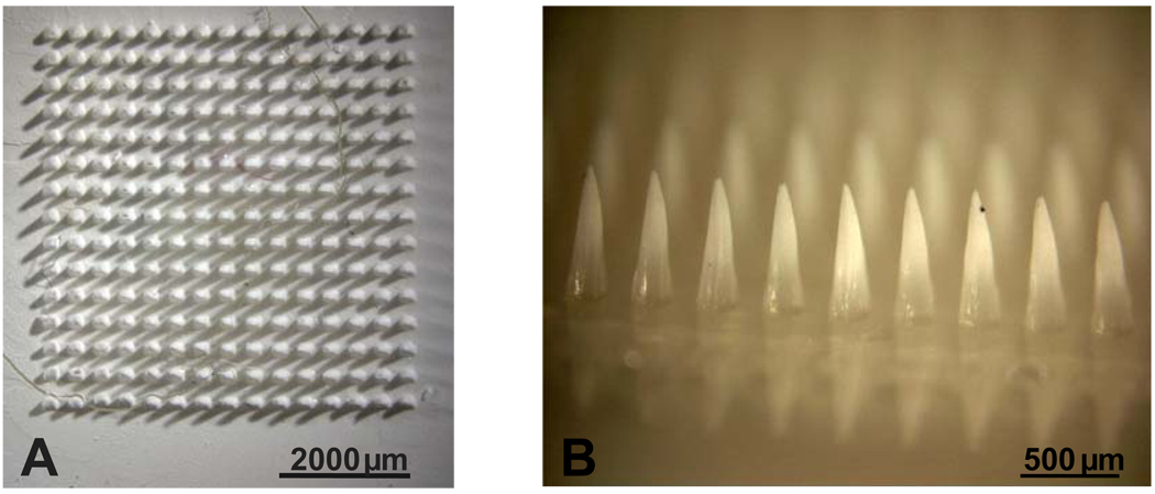
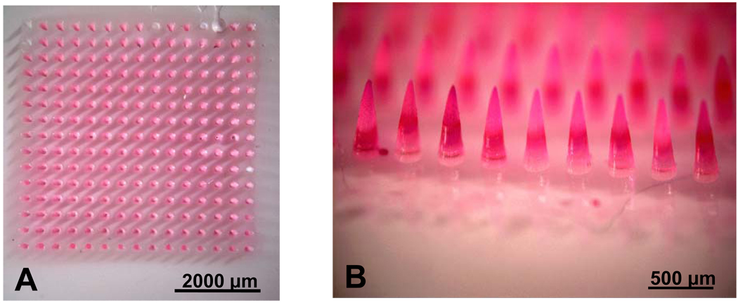
(A) Overhead view and (B) side view of pure PVP microneedles. (C) Overhead view and (D) side view of PVP polymer microneedles with sulforhodamine encapsulated within microneedles, but not in the base substrate. Each microneedle measures 750 µm in height, 250 µm in base diameter and 5 µm in tip radius.
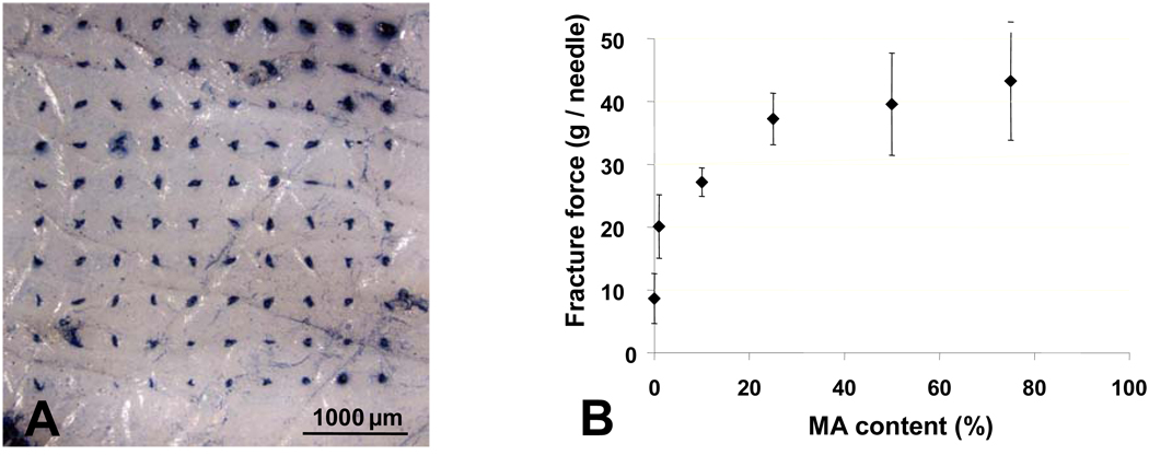
(A) Evidence of insertion of PVP polymer microneedles into porcine cadaver skin via skin marking test (B) The mechanical strength (fracture force) of copolymer PVP-MAA microneedles increases with increasing methacrylic acid (MAA) content.
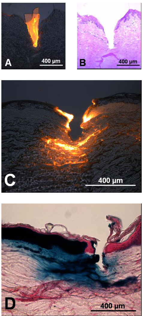
(A) Fluorescence microscopy image of a PVP polymer microneedle with encapsulated sulforhodamine inserted into porcine skin. (B) Brightfield microscopy image of the same skin section after microneedle removal showing the depth of microneedle insertion, stained with hemotoxilin and eosin. (C) Fluorescence microscopy image showing delivery of fluorescently labeled bovine serum albumin by PVP polymer microneedles to porcine skin. (D) Brightfield microscopy image of delivery of enzymatically active β-galactosidase via PVP polymer microneedles to porcine skin. The blue color represents the enzymatic conversion of X-gal by the delivered β-galactosidase
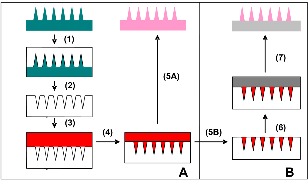
(A) (1) PDMS is poured onto microneedle master structure. (2) PDMS microneedle mold is cured and peeled off. (3) Liquid monomer and drug are pipetted onto the mold. (4) Vacuum is applied to pull the solution into the microneedle mold. (5A) System is placed under a UV lamp to polymerize microneedles, which are subsequently peeled out of the reusable mold. (B) (5B) Excess solution is removed from the surface. (6) A liquid monomer solution with no drug is applied to the surface. (7) System is placed under UV lamp to polymerize the microneedles, which are then peeled off.
References
-
- Walsh G. Nat Biotechnol. 2006;24:769. - PubMed
-
- Datamonitor. 2004:1. (Ed: D. USA)
-
- Langer R. Nature. 1998;392:5. - PubMed
-
- Prausnitz MR, Mitragotri S, Langer R. Nat Rev Drug Discov. 2004;3:115. - PubMed
-
- MR Prausnitz JM, Raeder-Devens J. In: Percutaneous Penetration Enhancers. Maibach ESaH., editor. Boca Raton: CRC Press; 2006.
Grants and funding
LinkOut - more resources
Full Text Sources
Other Literature Sources
