. 2012;7(12):e51008.
doi: 10.1371/journal.pone.0051008. Epub 2012 Dec 11.

Baicalin downregulates Porphyromonas gingivalis lipopolysaccharide-upregulated IL-6 and IL-8 expression in human oral keratinocytes by negative regulation of TLR signaling

Affiliations

Baicalin downregulates Porphyromonas gingivalis lipopolysaccharide-upregulated IL-6 and IL-8 expression in human oral keratinocytes by negative regulation of TLR signaling

Wei Luo et al. PLoS One. 2012.

Abstract

Periodontal (gum) disease is one of the main global oral health burdens and severe periodontal disease (periodontitis) is a leading cause of tooth loss in adults globally. It also increases the risk of cardiovascular disease and diabetes mellitus. Porphyromonas gingivalis lipopolysaccharide (LPS) is a key virulent attribute that significantly contributes to periodontal pathogenesis. Baicalin is a flavonoid from Scutellaria radix, an herb commonly used in traditional Chinese medicine for treating inflammatory diseases. The present study examined the modulatory effect of baicalin on P. gingivalis LPS-induced expression of IL-6 and IL-8 in human oral keratinocytes (HOKs). Cells were pre-treated with baicalin (0-80 µM) for 24 h, and subsequently treated with P. gingivalis LPS at 10 µg/ml with or without baicalin for 3 h. IL-6 and IL-8 transcripts and proteins were detected by real-time polymerase chain reaction and enzyme-linked immunosorbent assay, respectively. The expression of nuclear factor-κB (NF-κB), p38 mitogen-activated protein kinase (MAPK) and c-Jun N-terminal kinase (JNK) proteins was analyzed by western blot. A panel of genes related to toll-like receptor (TLR) signaling was examined by PCR array. We found that baicalin significantly downregulated P. gingivalis LPS-stimulated expression of IL-6 and IL-8, and inhibited P. gingivalis LPS-activated NF-κB, p38 MAPK and JNK. Furthermore, baicalin markedly downregulated P. gingivalis LPS-induced expression of genes associated with TLR signaling. In conclusion, the present study shows that baicalin may significantly downregulate P. gingivalis LPS-upregulated expression of IL-6 and IL-8 in HOKs via negative regulation of TLR signaling.


Conflict of interest statement

Competing Interests: The authors have declared that no competing interests exist.

Figures

Figure 1
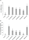
Figure 1. Baicalin significantly downregulates P. gingivalis LPS-upregulated IL-6 expression.
A. Baicalin (BI) at 40 µM and 80 µM significantly downregulated P. gingivalis (P.g.) LPS-upregulated IL-6 mRNA expression. B. Baicalin at 10 µM, 20 µM, 40 µM, and 80 µM markedly downregulated P.g. LPS-upregulated IL-6 protein expression. Cells treated with culture media alone served as the blank control group, and those treated with P.g. LPS (10 µg/ml) alone represented the positive control group. Cells treated with 0.08% DMSO and P.g. LPS at 10 µg/ml served as the vehicle control group. Data of three independent experiments were depicted as relative fold change as compared with the blank control group (set as 1) (A), or presented as protein concentration (B). *p<0.05 and **p<0.01 as compared with the positive control group (P.g. LPS).
Figure 2
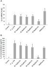
Figure 2. Baicalin significantly downregulates P. gingivalis LPS-upregulated IL-8 expression.
A. Baicalin (BI) at 80 µM significantly downregulated P. gingivalis (P.g.) LPS-upregulated IL-8 mRNA expression. B. Baicalin at 80 µM significantly downregulated P.g. LPS-upregulated IL-8 protein expression. Cells treated with culture media alone served as the blank control group, and those treated with P.g. LPS (10 µg/ml) alone represented the positive control group. Cells treated with 0.08% DMSO and P.g. LPS at 10 µg/ml served as the vehicle control group. Data of three independent experiments were depicted as relative fold change as compared to the blank control group (set as 1) (A), or presented as protein concentration (B). *p<0.01 as compared with the positive control group (P.g. LPS).
Figure 3
Figure 3. Baicalin inhibits P. gingivalis LPS-induced activation of NF-κB, p38 MAPK and JNK.
A. The representative western blot experiment was performed by pooling cytoplasmic protein extracts equally from three independent experiments. 25 µg aliquots were loaded into each lane. The membrane was firstly probed with the rabbit anti-phospho-IκBα mAbs (1∶2000), and sequentially stripped and re-probed with rabbit anti-phospho-p38 MAPK mAbs (1∶2000), rabbit anti-phospho-JNK mAbs (1∶2000), and rabbit anti-IκBα mAbs (1∶2000). For loading control, the membrane was probed with rabbit anti-α-tubulin mAbs (1∶4000). B. The densitometry analysis of the signals. Cells treated with culture media alone served as the blank control group, and those treated with P. gingivalis (P.g.) LPS (10 µg/ml) alone represented the positive control group. Cells treated with P.g. LPS at 10 µg/ml and 0.08% DMSO served as the vehicle control group. Data of three independent experiments were depicted as relative fold change as compared with the blank control group (set as 1). For the p-JNK protein, the positive control group (LPS) was set as 1 since the signals of the blank control group at 15 min and 30 min were undetectable. *p<0.05 and **p<0.01 as compared with the respective positive control group (LPS) at each time point. BI: baicain.
Figure 4
Figure 4. Baicalin suppresses P. gingivalis LPS-induced nuclear translocation of p65.
A. The representative experiment was performed by pooling nuclear protein extracts equally from three independent experiments. 2 µg aliquots were added to each well. The assay was carried out according to the manufacturer’s instruction. B. The intensity analysis of the luminescent signals. Cells treated with culture media alone served as the blank control group, and those treated with P. gingivalis (P.g.) LPS (10 µg/ml) alone represented the positive control group. Cells treated with P.g. LPS at 10 µg/ml and 0.08% DMSO served as the vehicle control group. Data from three independent experiments were depicted as relative fold change as compared with the blank control groups (set as 1). *p<0.05 and **p<0.01 as compared with the respective positive control group (LPS) at each time point. BI: baicalin.

References

    1. Jin LJ, Armitage GC, Klinge B, Lang NP, Tonetti M, et al. (2011) Global oral health inequalities: Task group-periodontal disease. Adv Dent Res 23: 221–226. - PubMed
    1. Parahitiyawa NB, Jin LJ, Leung WK, Yam WC, Samaranayake LP (2009) Microbiology of odontogenic bacteraemia: beyond endocarditis. Clin Microbiol Rev 22: 46–64. - PMC - PubMed
    1. Li X, Tse HF, Jin LJ (2011) Novel endothelial biomarkers: implications for periodontal disease and CVD. J Dent Res 90: 1062–1069. - PubMed
    1. Lalla E, Papapanou PN (2011) Diabetes mellitus and periodontitis: a tale of two common interrelated diseases. Nat Rev Endocrinol 7: 738–748. - PubMed
    1. Darveau RP (2010) Periodontitis: a polymicrobial disruption of host homeostasis. Nat Rev Microbiol 8: 481–490. - PubMed

Publication types

MeSH terms