. 2012;7(12):e51574.
doi: 10.1371/journal.pone.0051574. Epub 2012 Dec 11.

Depletion of resident macrophages does not alter sensory regeneration in the avian cochlea

Affiliations

Depletion of resident macrophages does not alter sensory regeneration in the avian cochlea

Mark E Warchol et al. PLoS One. 2012.

Abstract

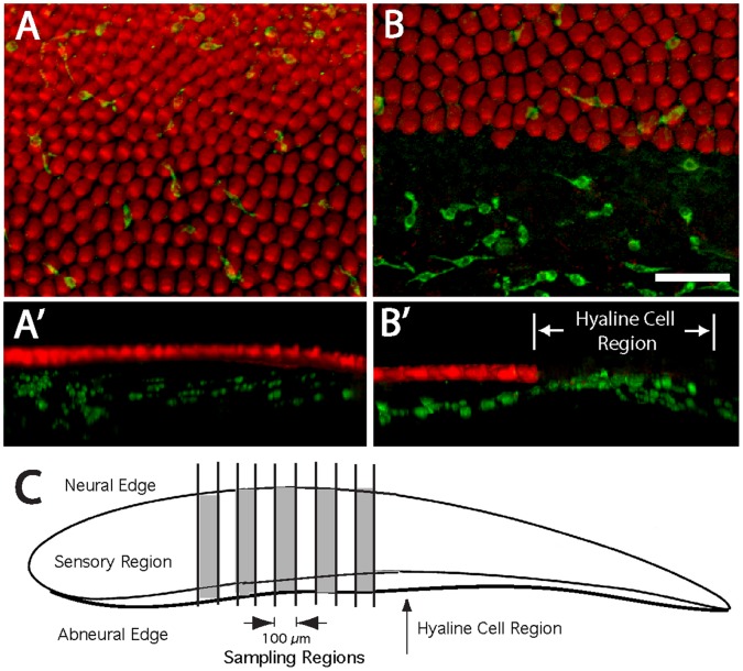
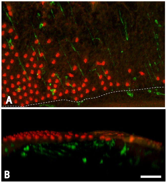
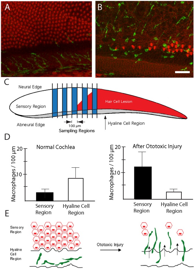
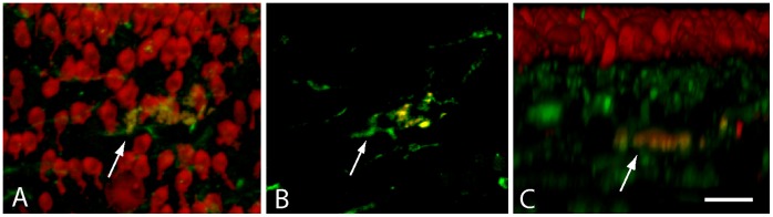
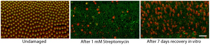
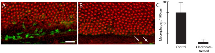
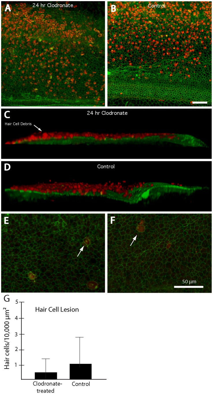
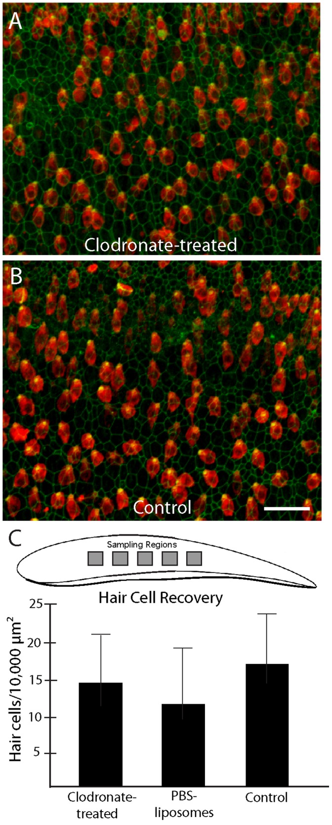
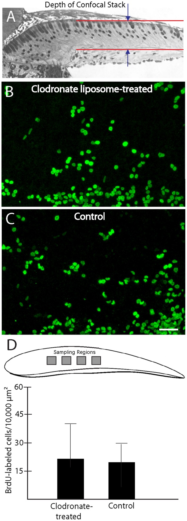
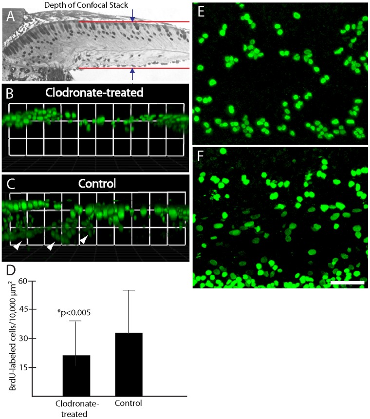
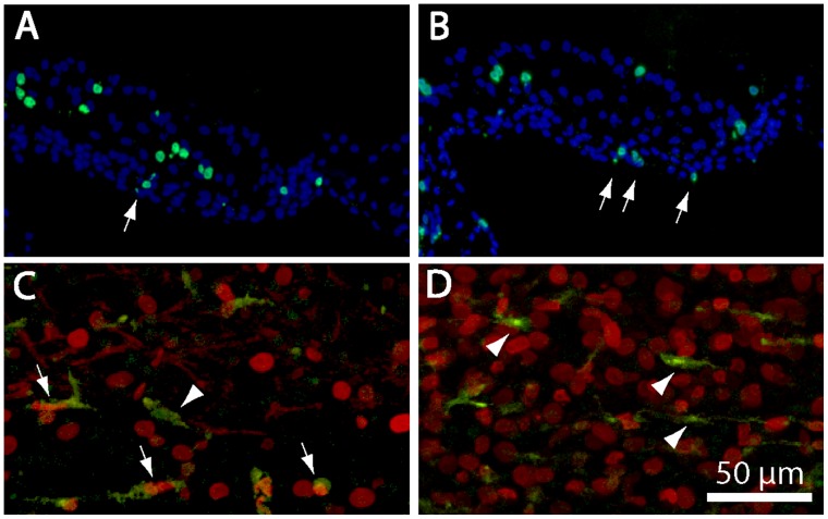
Macrophages are the primary effector cells of the innate immune system and are also activated in response to tissue injury. The avian cochlea contains a population of resident macrophages, but the precise function of those cells is not known. The present study characterized the behavior of cochlear macrophages after aminoglycoside ototoxicity and also examined the possible role of macrophages in sensory regeneration. We found that the undamaged chick cochlea contains a large resting population of macrophages that reside in the hyaline cell region, immediately outside the abneural (inferior) border of the sensory epithelium. Following ototoxic injury, macrophages appear to migrate out of the hyaline cell region and towards the basilar membrane, congregating immediately below the lesioned sensory epithelium. In order to determine whether recruited macrophages contribute to the regeneration of sensory receptors, we quantified supporting cell proliferation and hair cell recovery after the elimination of most resident macrophages via application of liposomally-encapsulated clodronate. Examination of macrophage-depleted specimens at two days following ototoxic injury revealed no deficits in hair cell clearance, when compared to normal controls. In addition, we found that elimination of macrophages did not affect either regenerative proliferation of supporting cells or the production of replacement hair cells. However, we did find that macrophage-depleted cochleae contained reduced numbers of proliferative mesothelial cells below the basilar membrane. Our data suggest that macrophages are not required for normal debris clearance and regeneration, but that they may play a role in the maintenance of the basilar membrane.


Conflict of interest statement

Competing Interests: The authors have declared that no competing interests exist.

Figures

Figure 1
Figure 1. Macrophages in the uninjured chick cochlea.
Surface views revealed macrophages (green) associated with the sensory region (A). In addition, numerous macrophages were present in the hyaline/cuboidal cell region, which is immediately outside the inferior border of the sensory epithelium (B). Three-dimensional reconstructions of confocal image stacks indicated that macrophages in the sensory region were mainly located below the sensory epithelium (A′, B′). Macrophages in the sensory and nonsensory (hyaline cell) regions of the cochlea were quantified from five 100 µm-wide strips, located in the midregion of the cochlea (shaded areas, C). Green: KUL01 (macrophages), Red: phalloidin. Scale bar = 30 µm.
Figure 2
Figure 2. Response of cochlear macrophages to ototoxic injury.
Chicks received three injections of streptomycin (1,200 mg/kg, 1/day), which killed most hair cells in the proximal-most ∼50% of the sensory region. Cochleae were fixed 24 hr after the final injection, and macrophages were identified by immunoreactivity for KUL01 (green). Hair cell stereocilia and cell-cell junctions were also labeled with phalloidin (red). Surface views (A) indicated that the sensory regions of lesioned cochlea contained increased numbers of macrophages. The dashed line indicates the inferior border of the sensory epithelium. Examination of three dimensional renderings of the confocal image stacks (B) indicated that most newly-recruited macrophages remained below the sensory epithelium. Scale bar = 30 µm.
Figure 3
Figure 3. Redistribution of cochlear macrophages following ototoxic injury.
The hyaline cell regions of undamaged cochleae contained numerous macrophages (A, green). After ototoxic injury, increased numbers of macrophages were observed within the sensory region (B). Macrophage numbers in the sensory and hyaline regions were quantified using the scheme shown in (C). Data from undamaged cochleae indicated that the hyaline cell region contained more macrophages than did the adjacent sensory region (D, left, p<0.001). After injury, however, macrophages in the sensory region outnumbered those in the hyaline region (D, right, p<0.001). This apparent redistribution suggests that resting macrophages migrate into the sensory region in response to hair cell injury, as schematized in (E). Scale bar = 30 µm.
Figure 4
Figure 4. Co-localization of macrophages with hair cell debris.
Cochleae were fixed at one day after completion of streptomycin injections (see text) and immunolabeled for KUL01 (green, macrophages) and myosin VIIA (red, hair cells). Specimens contained a small number of cells that were double-labeled for both markers (arrows). Such labeling is suggestive of macrophages having phagocytosed the remains of injured hair cells. Reconstruction of confocal image stacks revealed that such cells were below the sensory epithelium (C). Scale bar = 30 µm.
Figure 5
Figure 5. Ototoxic injury and hair cell regeneration in organotypic culture.
Cultures of chick cochleae were treated for 24 hr with 1 mM streptomycin, which killed nearly all hair cells in the sensory epithelium (center). When such lesioned specimens were maintained for an additional seven days in streptomycin-free medium, significant hair cell recovery was observed (right). All images show the midregion of the chick cochlea. Labels: red-Myosin VIIA (hair cells), green-phalloidin. Scale bar = 30 µm.
Figure 6
Figure 6. Treatment with liposomally-encapsulated clodronate reduces the number of macrophages in cochlear cultures.
(A) Untreated cochlear cultures possess numerous macrophages (green), particularly in the hyaline/cuboidal cell region. (B) After treatment for 24 hr in clodronate-containing liposomes, very few macrophages remain (arrows). In contrast, hair cell morphology did not appear to be affected by clodronate treatment (red: phalloidin). (C) Quantification of macrophages in clodronate-treated vs. control cultures (n = four samples/specimen from eight clodronate-treated and six control cochleae) demonstrates a sharp reduction in the resident macrophage population after clodronate treatment (p<0.001). Scale bar = 30 µm.
Figure 7
Figure 7. Elimination of macrophages does not affect the clearance of hair cell debris after ototoxic injury.
(A, B) Surface views of cochleae fixed at 48 hr after streptomycin treatment. High levels of hair cell debris (labels: red-myosin VIIA; green-phalloidin) were present in all specimens, regardless of pretreatment. (C, D) Three-dimensional reconstructions of confocal image stacks indicated that nearly all hair cell debris was located above the lumenal surface of the sensory epithelium. (E, F) In contrast, confocal images that were confined to the sensory region revealed very low levels of hair cell debris remaining in the epithelia of either clodronate-treated (E) or control (F) cultures. Arrows in (E) and (F) point to myosin VIIA-labeled cells that remained within the sensory epithelium at 48 hours after ototoxic injury. Quantification of such cells confirms that macrophage depletion did not impair the removal of hair cell debris from the sensory epithelium (G). Scale bars = 50 µm.
Figure 8
Figure 8. Depletion of cochlear macrophages does not affect hair cell recovery after ototoxic lesion.
Cochlea were treated with clodronate-containing or ‘empty’ liposomes, or received no pretreatment. They were then incubated for 24 hr in 1 mM streptomycin (to kill hair cells) and allowed to recover in vitro for an additional seven days. Nearly identical levels of hair cell recovery were observed in all specimens, regardless of pretreatment (A, B). Quantification of hair cell density from five 100×100 µm regions/cochlea confirmed that macrophage depletion had no effect on hair cell regeneration (C). Scale bar = 30 µm.
Figure 9
Figure 9. Macrophage depletion does not affect regenerative proliferation.
Cochleae were treated for 24 hr. in clodronate-containing or control medium, followed by 24 hr in 1 mM streptomycin. Specimens were then rinsed and maintained for an additional four days in medium that contained BrdU. Confocal microscopy was used to image BrdU-labeled cells within the sensory epithelia of the cochleae (A). Similar patterns of BrdU labeling were observed in clodronate-treated (B) and control (C) cochlea. Quantification of BrdU labeled cells (from four 100×100 µm regions/specimen) confirmed that supporting cell proliferation was not affected by clodronate treatment and macrophage depletion (D, p = 0.28). Scale bar = 30 µm.
Figure 10
Figure 10. Clodronate treatment reduces proliferation of nonsensory cells in the chick cochlea.
(A) Confocal image stacks of BrdU-labeled cells (green) in clodronate-treated and control specimens were obtained by scanning through the entire specimen (including both the sensory epithelium and the underlying basilar membrane region). (B, C) Three-dimensional reconstructions of those images indicated that clodronate treatment selectively reduced proliferation of cells below the sensory epithelium (C, arrowheads). Surface views of clodronate-treated (E) and control (F) cultures also suggested fewer BrdU-labeled cells after clodronate treatment. Quantification of all BrdU-labeled cells in those specimens confirmed that clodronate reduces proliferation (p<0.005). Scale bar = 30 µm.
Figure 11
Figure 11. Location and identity of BrdU-labeled cells in cochlear cultures.
Frozen sections of cultured cochleae typically contained BrdU-labeled cells (green) in the sensory epithelium and within the basilar membrane (arrows, A, B). Cell nuclei are labeled with DAPI (blue). (C, D) Confocal images of cochleae that were double labeled for KUL01 (green, macrophages) and BrdU (red) indicate that macrophages constituted a minority of proliferating cells (arrows). Numerous quiescent macrophages (arrow heads) were also present. Scale bar (for images C and D) = 50 µm.

Similar articles

Cited by

References

    1. Rosenblatt J, Raff MC, Cramer LP (2001) An epithelial cell destined for apoptosis signals its neighbors to extrude it by an actin- and myosin-dependent mechanism. Curr Biol 11: 1847–1857. - PubMed
    1. Hirose K, Westrum LE, Cunningham DE, Rubel EW (2004) Electron microscopy of degenerative changes in the chick basilar papilla after gentamicin exposure. J Comp Neurol 470: 164–180. - PubMed
    1. Bird JE, Daudet N, Warchol ME, Gale JE (2010) Rapid elimination of dying sensory hair cells maintains epithelial integrity in the avian inner ear. J Neurosci 30: 12545–12556. - PMC - PubMed
    1. Warchol ME (1997) Macrophage activity in the avian cochlea: demonstration of a resident population and recruitment to sites of hair cell lesions. J Neurobiol 33: 724–734. - PubMed
    1. Warchol ME (1999) Immune cytokines and dexamethasone influence sensory regeneration in the avian vestibular periphery. J Neurocytol 28: 889–900. - PubMed

Publication types

Substances