. 2012;7(12):e51798.
doi: 10.1371/journal.pone.0051798. Epub 2012 Dec 11.

Restriction of neural precursor ability to respond to Nurr1 by early regional specification

Affiliations

Restriction of neural precursor ability to respond to Nurr1 by early regional specification

Chiara Soldati et al. PLoS One. 2012.

Abstract

During neural development, spatially regulated expression of specific transcription factors is crucial for central nervous system (CNS) regionalization, generation of neural precursors (NPs) and subsequent differentiation of specific cell types within defined regions. A critical role in dopaminergic differentiation in the midbrain (MB) has been assigned to the transcription factor Nurr1. Nurr1 controls the expression of key genes involved in dopamine (DA) neurotransmission, e.g. tyrosine hydroxylase (TH) and the DA transporter (DAT), and promotes the dopaminergic phenotype in embryonic stem cells. We investigated whether cells derived from different areas of the mouse CNS could be directed to differentiate into dopaminergic neurons in vitro by forced expression of the transcription factor Nurr1. We show that Nurr1 overexpression can promote dopaminergic cell fate specification only in NPs obtained from E13.5 ganglionic eminence (GE) and MB, but not in NPs isolated from E13.5 cortex (CTX) and spinal cord (SC) or from the adult subventricular zone (SVZ). Confirming previous studies, we also show that Nurr1 overexpression can increase the generation of TH-positive neurons in mouse embryonic stem cells. These data show that Nurr1 ability to induce a dopaminergic phenotype becomes restricted during CNS development and is critically dependent on the region of NPs derivation. Our results suggest that the plasticity of NPs and their ability to activate a dopaminergic differentiation program in response to Nurr1 is regulated during early stages of neurogenesis, possibly through mechanisms controlling CNS regionalization.


Conflict of interest statement

Competing Interests: The authors have declared that no competing interests exist.

Figures

Figure 1
Figure 1. Immunodetection of Nestin in NPs lines derived from different brain regions and RT-PCR detection of regionally expressed genes.
A) Immunostaining for nestin (green) on NPs derived from a) cortex (CTX), b) lateral ganglionic eminence (GE), c) spinal cord (SC), d) midbrain (MB), e) subventricular zone (SVZ), f) mouse blastocyst (ES-D3). Nuclei were stained with DAPI (blue). Scale bar  = 20 μm. B). B) Real time PCR quantification of Ngn2, HoxB6, HoxB9, Dlx2, Six3 and FoxG1 expression in CTX-, GE- and SC-derived NPs cultured in proliferating conditions. Results are shown as the mean of the ratio between gene expression in NPs and in dissected E13.5 tissue (telencephalon or spinal cord) in four biological replicates. Error bars show the standard error of the mean. * P<0,05 ***p<0.001.
Figure 2
Figure 2. Nurr1 overexpression in NPs lines following lentivurs infection.
Nurr1 mRNA levels detected by Real Time PCR in CTX, GE, SC, MB, adult SVZ and in ES-D3 derived NPs in control conditions and after infection with Nurr1 lentivirus. Nurr1 mRNA level in E12 mouse embryo MB (E MB) is shown as the positive control. The error bars represent standard deviation (n = 3). *** P<0,001.
Figure 3
Figure 3. Evaluation of neuronal and glial cells in differentiated NPs lines.
A) Immunostaining for beta-III-tubulin (green) GFAP (red) on differentiated cells in control conditions (A, C, E, G, I.M) and after infection with Nurr1 lentivirus (B, D, F, H, L, N). (A, B) CTX, (C, D) GE, (E, F) SC, (G, H) MB, (I, L) adult SVZ, (M, N) ES-D3. Nuclei were stained with DAPI. B) Percentage of beta-III-tubulin and GFAP positive cells after differentiation of NPs. The reported values are the mean ± SEM of 10 observations from two independent experiments Scale bar  = 20 μm. The error bars represent standard deviation.
Figure 4
Figure 4. Induction of TH expression by Nurr1 overexpression: RT-PCR analysis.
TH mRNA level detected by Real Time PCR in differentiated CTX, GE, MB and ES-D3 NPs and in cells infected with Nurr1. TH mRNA was not detectable in other analyzed NPs. TH mRNA level in mouse MB (E MB) is reported as positive control. The error bars represent standard deviation (n = 3). *** P<0,001.
Figure 5
Figure 5. Induction of TH expression by Nurr1 overexpression: immunocytochemistry analysis.
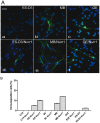
A) Immunostaining for TH (green) on differentiated cells in control conditions and after infection with Nurr1 lentivirus. Nuclei were stained with DAPI (blue). (a) ES-D3, (d) Nurr1 infected ES-D3, (b) MB, (e) MB Nurr1 infected (c) GE, (f) Nurr1 infected GE. B) Percentage of TH positive cells (vs total cell number). Scale bar  = 30 μm.
Figure 6
Figure 6. Activation of the dopaminergic markers DAT and Ptx3 by Nurr1 overexpression.
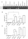
A) Representative samples of RT-PCR amplified products for DAT B) and C). real-time PCR of DAT (B) and Ptx3 (C) mRNA on differentiated cells in control conditions and after Nurr1 overexpression HPRT was used as normalizing gene. The error bars represent standard deviation (n = 3). ** P<0,02.

References

    1. Edlund T, Jessell TM (1999) Progression from extrinsic to intrinsic signaling in cell fate specification: a view from the nervous system. Cell 96: 211–224. - PubMed
    1. Ruiz I, Altaba A (1992) Planar and vertical signals in the induction and patterning of the Xenopus nervous system. Development 115: 67–80. - PubMed
    1. Bertrand N, Castro DS, Guillemot F (2002) Proneural genes and the specification of neural cell types. Nat Rev Neurosci 3: 517–30. - PubMed
    1. Petryniak MA, Potter GB, Rowitch DH, Rubenstein JL (2007) Dlx1 and Dlx2 control neuronal versus oligodendroglial cell fate acquisition in the developing forebrain. Neuron 55: 417–33. - PMC - PubMed
    1. Kim EJ, Battiste J, Nakagawa Y, Johnson JE (2008) Ascl1 (Mash1) lineage cells contribute to discrete cell populations in CNS architecture. Mol Cell Neurosci 38: 595–606. - PMC - PubMed

Publication types

MeSH terms

Substances