. 2013 May;125(3):430-45.
doi: 10.1111/jnc.12130. Epub 2013 Jan 7.

Deletion or activation of the aryl hydrocarbon receptor alters adult hippocampal neurogenesis and contextual fear memory

Affiliations

Deletion or activation of the aryl hydrocarbon receptor alters adult hippocampal neurogenesis and contextual fear memory

Sarah E Latchney et al. J Neurochem. 2013 May.

Abstract

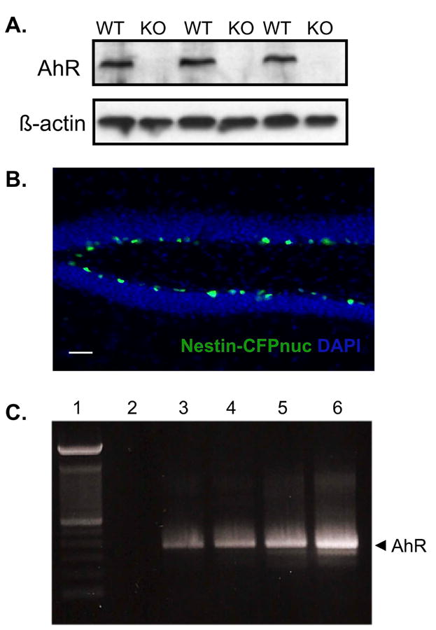
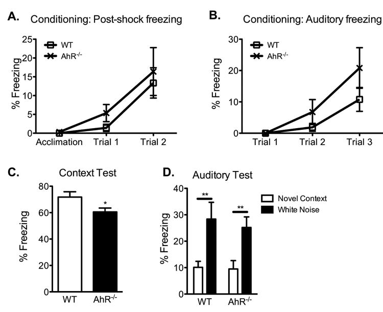
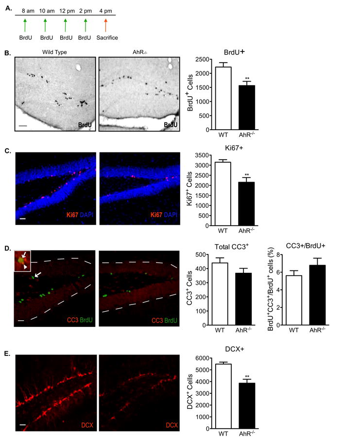
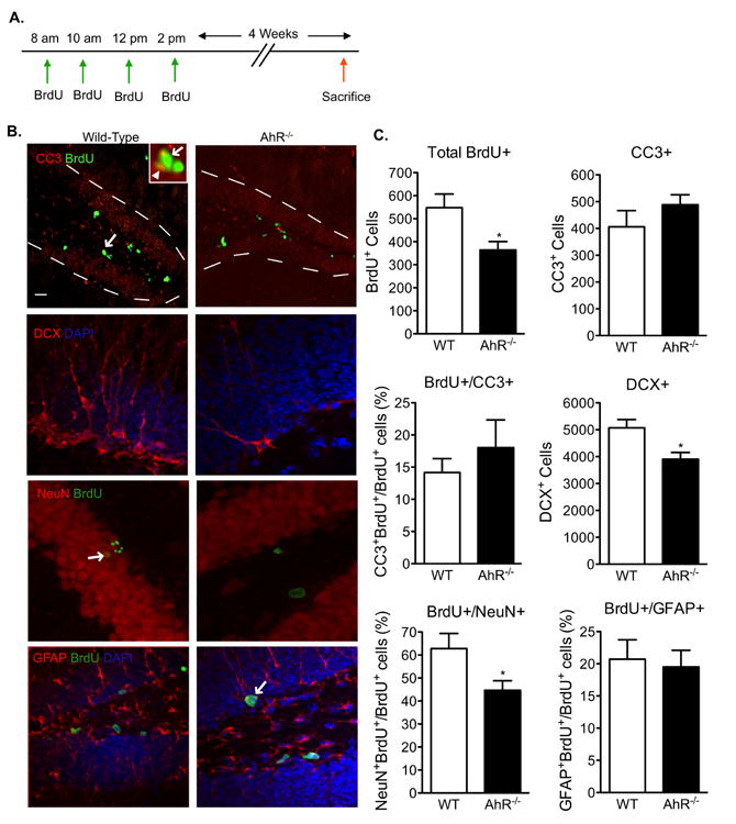
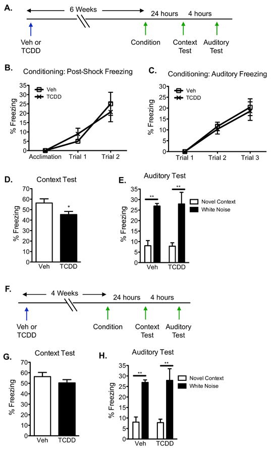
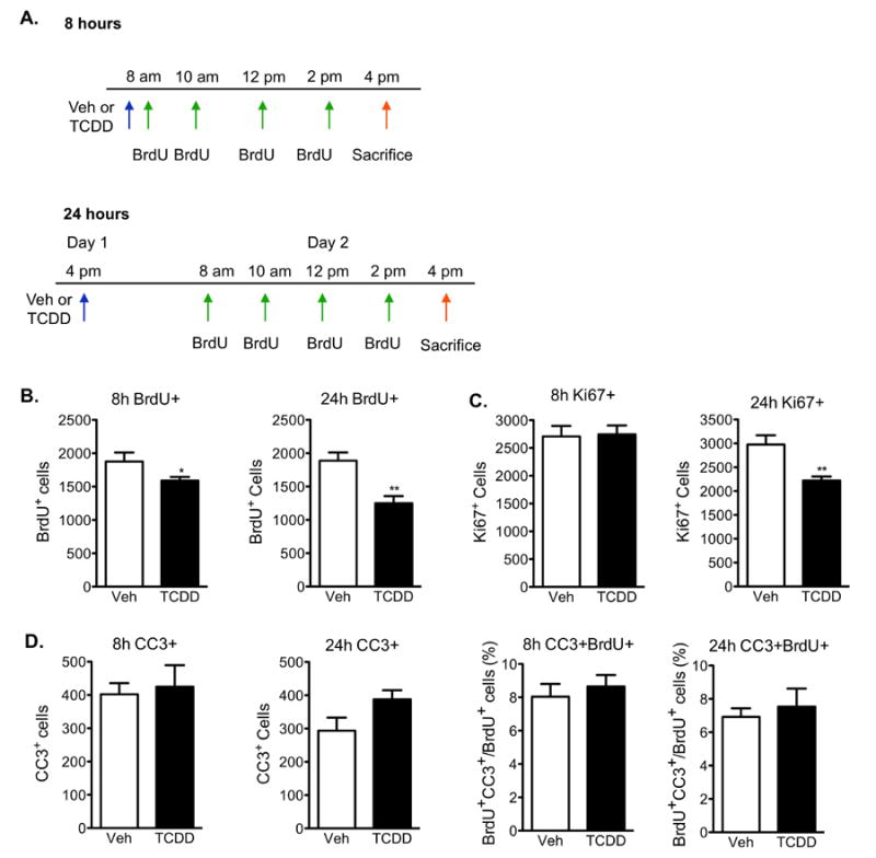
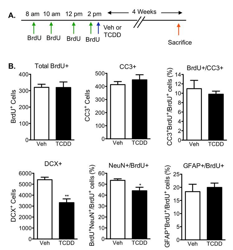
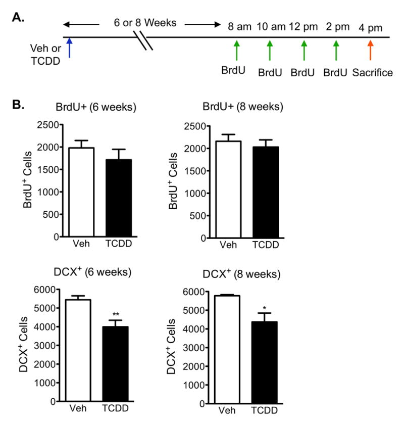
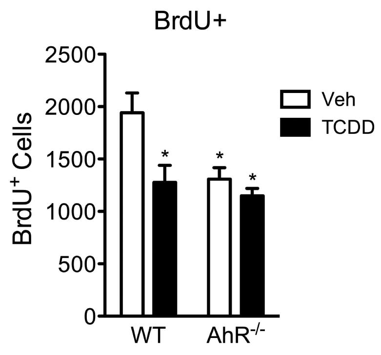
The aryl hydrocarbon receptor (AhR) is a ligand-activated transcription factor that mediates the toxicity of dioxin and serves multiple developmental roles. In the adult brain, while we now localize AhR mRNA to nestin-expressing neural progenitor cells in the dentate gyrus (DG) of the hippocampus, its function is unknown. This study tested the hypothesis that AhR participates in hippocampal neurogenesis and associated functions. AhR deletion and activation by the potent environmental toxicant, 2,3,7,8-tetrachlorodibenzo-p-dioxin (TCDD), adversely impacted neurogenesis and cognition. Adult AhR-deficient mice exhibited impaired hippocampal-dependent contextual fear memory while hippocampal-independent memory remained intact. AhR-deficient mice displayed reduced cell birth, decreased cell survival, and diminished neuronal differentiation in the DG. Following TCDD exposure, wild-type mice exhibited impaired hippocampal-dependent contextual memory, decreased cell birth, reduced neuronal differentiation, and fewer mature neurons in the DG. Glial differentiation and apoptosis were not altered in either TCDD-exposed or AhR-deficient mice. Finally, defects observed in TCDD-exposed mice were dependent on AhR, as TCDD had no negative effects in AhR-deficient mice. Our findings suggest that AhR should be further evaluated as a potential transcriptional regulator of hippocampal neurogenesis and function, although other sites of action may also warrant consideration. Moreover, TCDD exposure should be considered as an environmental risk factor that disrupts adult neurogenesis and potentially related memory processes.


Conflict of interest statement

No conflicts of interests exist.

Figures

Figure 1
Figure 1. Nestin-positive neural progenitor cells in the adult dentate gyrus express AhR
(A)Protein lysates were isolated from the hippocampus of 3 adult wild-type (WT) and 3 adult AhR knockout (KO) mice. AhR protein content (10μg) was analyzed by immunoblotting. The corresponding β-actin blot served as a loading control. (B) Representative image of the dentate gyrus from an adult nestin-CFPnuc mouse. CFP-positive cells (green) were restricted to the SGZ of the dentate gyrus. Scale bar=100μm. (C) Representative gel from RT-PCR analysis for AhR mRNA. Lane 1: 100bp DNA ladder, Lane 2: Negative control (non-reverse transcribed mRNA). Lanes 3-5: 3 independent preparations of CFP sorted cells from nestin-CFPnuc mice. Lane 6: Positive control (mRNA isolated from C17.2 cells).
Figure 2
Figure 2. AhR deletion impairs hippocampal-dependent memory
(A and B) Mean percent freezing during the acquisition, post-shock intervals, and auditory stimuli during conditioning increased with repeated pairing but did not differ between wild-type and AhR-/- mice (N=10 mice/genotype; 2-way ANOVA). (C) In the hippocampal-dependent context test, AhR-/- mice show reduced freezing to the conditioned context compared to age-matched wild-type animals (N=10 mice/genotype; Student's t-test, * p<0.05). (D) Both wild-type and AhR-/- mice showed low freezing to a novel context and elevated freezing to the conditioned white noise stimulus (N=10 mice/genotype; 2-way ANOVA with Bonferroni post-hoc test, ** p<0.01). Results are expressed as means ± S.E.M.
Figure 3
Figure 3. Reduced cell proliferation and neuronal differentiation in the dentate gyrus of AhR-deficient mice
(A)Paradigm used to examine neural progenitor cell birth in wild-type and AhR-/- mice. Representative images and quantitative analysis of (B) BrdU, (C) Ki67, (D) Cleaved Caspase-3 (CC3) and CC3-BrdU double positive, and (E) doublecortin (DCX) positive cells in the dentate gyrus of wild-type and AhR-/- mice. Scale bar=100μm in (B) and 500μm in (C-E). The arrow in the low magnification image points to a double-positive BrdU+/CC3+ cell. The inset is a higher magnification image of the double-positive cell (arrow in inset points to the BrdU+ green signal and arrowhead points to the CC3+ red signal). Data represents means ± S.E.M. (N=10 mice/genotype); Student's t-test (* p<0.05, ** p < 0.01).
Figure 4
Figure 4. Cell survival and neuronal differentiation is reduced in AhR-/- mice
(A)Paradigm used to examine the survival and differentiation of newly born cells in AhR-/- and WT mice 4 weeks after BrdU labeling. (B) Representative images of the dentate gyrus stained for BrdU/CC-3, DCX, NeuN/BrdU, and GFAP/BrdU. Overlays are shown. Scale bar=100μm for BrdU/CC3 image and 10μm for all others. The arrow in the BrdU/CC3 low magnification image points to a to double-positive BrdU+/CC3+ cell. The inset is a higher magnification image of the double-positive cell (arrow in inset points to the BrdU+ green signal and arrowhead pionts to the CC3+ red signal). The arrow in the BrdU/NeuN and BrdU/GFAP images points to a double-positive cell. (C) Quantification revealed that cell survival (total BrdU+ cells) was reduced whereas apoptosis (CC-3) was not altered in AhR-/- mice. Early (DCX+ cells) and late (NeuN- and BrdU-double positive cells) neuronal differentiation was reduced in AhR-/- mice. Maturation of newborn cells into GFAP+ astrocytes was not affected. Results are expressed as means ± S.E.M. (N=5 mice/group); Student's t-test (* p<0.05).
Figure 5
Figure 5. TCDD exposure leads to hippocampal-dependent memory impairment
(A)Contextual fear conditioning paradigm to test for hippocampal-dependent and independent function. (B and C) Mean percent freezing during the acquisition, post-shock intervals, and auditory stimuli during conditioning increased with repeated pairing but did not differ between vehicle and TCDD exposed mice (N=10 mice/genotype; 2-way ANOVA). (C) In the hippocampal-dependent context test, TCDD exposed mice froze less to the conditioned context compared to vehicle exposed animals (N=10 mice/genotype; Student's t-test, * p<0.05). (D) All mice showed low freezing to a novel context and elevated freezing to the conditioned white noise stimulus (N=10 mice/genotype; 2-way ANOVA with Bonferroni post-hoc tests, ** p<0.01). Results are expressed as means ± S.E.M.
Figure 6
Figure 6. TCDD reduces cell proliferation in adult hippocampus without inducing cell death
(A)Paradigm used to investigate neural cell proliferation in the dentate gyrus 8h and 24h following TCDD exposure. (B) Quantification of BrdU+ cells in the SGZ demonstrated a reduction in S-phase entry 8h after TCDD exposure compared to vehicle-treated animals, which became more pronounced at 24h. (C) Quantification of Ki67-positive cells revealed a significant reduction in newborn cells in all phases of the cell cycle following 24h exposure to TCDD, but not at 8h. (D) Quantification of CC3-single and CC3- and BrdU-double positive cells relative to total number of BrdU-positive cells in the SGZ demonstrated that TCDD does not induce apoptosis at either timepoint. All results are expressed as means ± S.E.M. (N=10 mice/group); Student's t-test (* p<0.05, ** p <0.01).
Figure 7
Figure 7. Neuronal, but not glial, differentiation is reduced 4 weeks following TCDD exposure
(A)Paradigm used to examine the survival and differentiation of newly born cells 4 weeks following TCDD exposure. (B) Quantification revealed that cell survival (total BrdU+ cells) and cell death (CC-3) were not affected with TCDD exposure. Early (DCX+ cells) and late (NeuN- and BrdU-double positive cells) neuronal differentiation was reduced with TCDD exposure. Maturation of newborn cells into GFAP+ astrocytes was not affected with TCDD exposure. Results are expressed as means ± S.E.M. (N=5 mice/group); Student's t-test (* p<0.05, ** p <0.01).
Figure 8
Figure 8. Sustained reduction in neuronal differentiation, but not cell birth, 6 and 8 weeks post TCDD exposure
(A)Paradigm used to analyze long-term effects on neural cell birth and differentiation following TCDD exposure. (B) Quantification of BrdU+ cells in the SGZ demonstrated no significant changes in cell birth 6 and 8 weeks after TCDD exposure compared to vehicle-exposed mice. (C) Quantification of DCX+ cells in the SGZ demonstrated a persistent reduction in DCX expression 6 and 8 weeks after TCDD exposure. Results are expressed as means ± S.E.M. (N=5 mice/group); Student's t-test (* p<0.05, ** p <0.01).
Figure 9
Figure 9. AhR-deficient mice are resistant to TCDD-induced neurotoxicity
Quantification of BrdU+ cells in the SGZ revealed no difference in BrdU-positive cells between vehicle- and TCDD-exposed AhR-/- mice, indicating TCDD neurotoxicity requires the AhR. Results are expressed as means ± S.E.M. (N=3 mice/group); 2-way ANOVA with Bonferroni post-hoc tests (* p<0.05 versus vehicle-exposed wild-types).

Similar articles

Cited by

References

    1. Aasebo IE, Blankvoort S, Tashiro A. Critical maturational period of new neurons in adult dentate gyrus for their involvement in memory formation. Eur J Neurosci. 2011;33:1094–1100. - PubMed
    1. Ables JL, Decarolis NA, Johnson MA, Rivera PD, Gao Z, Cooper DC, Radtke F, Hsieh J, Eisch AJ. Notch1 is required for maintenance of the reservoir of adult hippocampal stem cells. J Neurosci. 2010;30:10484–10492. - PMC - PubMed
    1. Anderson ML, Sisti HM, Curlik DM, 2nd, Shors TJ. Associative learning increases adult neurogenesis during a critical period. Eur J Neurosci. 2011;33:175–181. - PMC - PubMed
    1. Baader SL, Sanlioglu S, Berrebi AS, Parker-Thornburg J, Oberdick J. Ectopic overexpression of engrailed-2 in cerebellar Purkinje cells causes restricted cell loss and retarded external germinal layer development at lobule junctions. J Neurosci. 1998;18:1763–1773. - PMC - PubMed
    1. Baader SL, Vogel MW, Sanlioglu S, Zhang X, Oberdick J. Selective disruption of “late onset” sagittal banding patterns by ectopic expression of engrailed-2 in cerebellar Purkinje cells. J Neurosci. 1999;19:5370–5379. - PMC - PubMed

Publication types

MeSH terms