

Effects of placement angle and direction of orthopedic force application on the stability of orthodontic miniscrews
- PMID: 23241005
- PMCID: PMC8754024
- DOI: 10.2319/090112-703.1
Effects of placement angle and direction of orthopedic force application on the stability of orthodontic miniscrews
Abstract
Objectives: To evaluate the influence of placement angle and direction of orthopedic force application on the stability of miniscrews.
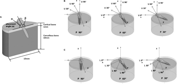
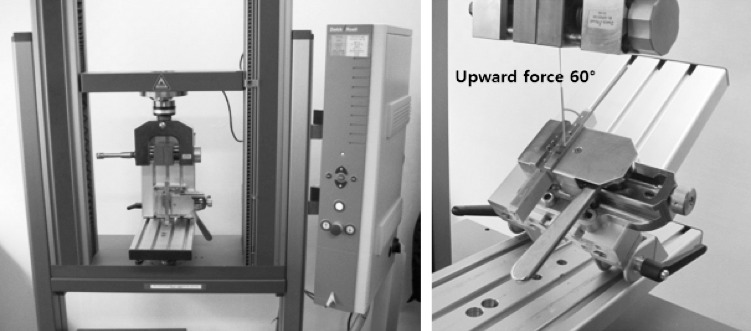
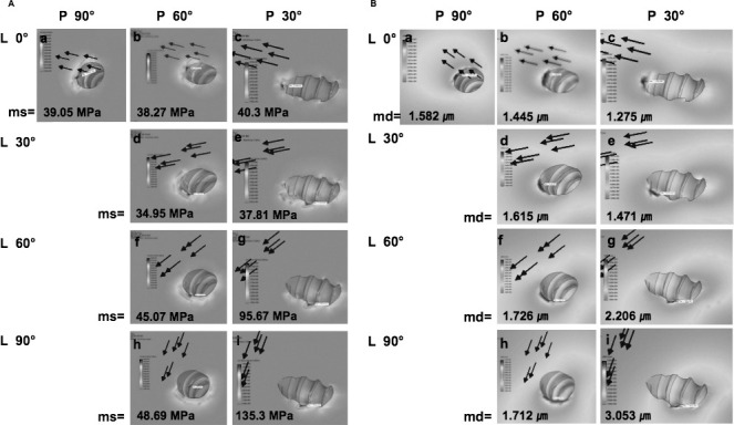
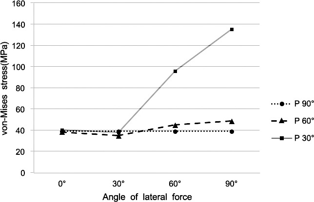
Materials and methods: Finite element analysis was performed using miniscrews inserted into supporting bone at angles of 90°, 60°, and 30° (P90°, P60°, and P30°). An orthopedic heavy force of 800 gf was applied to the heads of the miniscrews in four upward (U0°, U30°, U60°, U90°) or lateral (L0°, L30°, L60°, L90°) directions. In addition, pull-out strength of the miniscrews was measured with various force directions and cortical bone thicknesses.
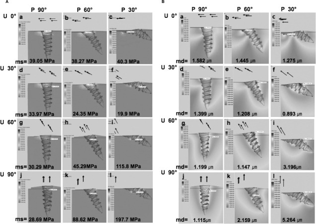
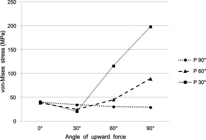
Results: Miniscrews with a placement angle of 30° (P30°) and 60° (P60°) showed a significant increase in maximum von Mises stress following the increase in lateral force vectors (U30°, U60°, U90°) compared to those with a placement angle of 90° (P90°). In accordance, the pull-out strength was higher with the axial upward force when compared to the upward force with lateral vectors. Maximum von Mises stress and displacement of the miniscrew increased as the angle of lateral force increased (L30°, L60°, L90°). However, a more dramatic increase in maximum von Mises stress was noted in P30° than in P60° and P90°.
Conclusion: Placement of the miniscrew perpendicular to the cortical bone is advantageous in terms of biomechanical stability. Placement angles of less than 60° can reduce the stability of miniscrews when orthopedic forces are applied in various directions.
Figures
Similar articles
-
Finite element analysis of miniscrew placement in mandibular alveolar bone with varied angulations.Eur J Orthod. 2015 Feb;37(1):56-9. doi: 10.1093/ejo/cju006. Epub 2014 Aug 1. Eur J Orthod. 2015. PMID: 25086029
-
Changes in stress distribution of orthodontic miniscrews and surrounding bone evaluated by 3-dimensional finite element analysis.Am J Orthod Dentofacial Orthop. 2011 Dec;140(6):e273-80. doi: 10.1016/j.ajodo.2011.06.025. Am J Orthod Dentofacial Orthop. 2011. PMID: 22133961
-
The ideal insertion angle after immediate loading in Jeil, Storm, and Thunder miniscrews: A 3D-FEM study.Int Orthod. 2020 Sep;18(3):503-508. doi: 10.1016/j.ortho.2020.03.003. Epub 2020 May 6. Int Orthod. 2020. PMID: 32387220
-
Effects of mechanical vibration on miniscrew implants and bone: Fem analysis.Int Orthod. 2019 Mar;17(1):38-44. doi: 10.1016/j.ortho.2019.01.022. Epub 2019 Feb 13. Int Orthod. 2019. PMID: 30770332
-
Effect of cortical bone thickness on shear stress and force in orthodontic miniscrew-bone interface - A finite element analysis.Biomed Phys Eng Express. 2024 Jul 19;10(5). doi: 10.1088/2057-1976/ad6160. Biomed Phys Eng Express. 2024. PMID: 38986445
Cited by
-
Effect of miniscrew insertion angle in the maxillary buccal plate on its clinical survival: a randomized clinical trial.Prog Orthod. 2021 Aug 2;22(1):22. doi: 10.1186/s40510-021-00370-8. Prog Orthod. 2021. PMID: 34337677 Free PMC article. Clinical Trial.
-
Anchoring structure of the calvarial periosteum revealed by focused ion beam/scanning electron microscope tomography.Sci Rep. 2015 Dec 2;5:17511. doi: 10.1038/srep17511. Sci Rep. 2015. PMID: 26627533 Free PMC article.
-
Evaluation of stress generation on the cortical bone and the palatal micro-implant complex during the implant-supported en masse retraction in lingual orthodontic technique using the FEM: Original research.J Dent Res Dent Clin Dent Prospects. 2019 Summer;13(3):192-199. doi: 10.15171/joddd.2019.030. Epub 2019 Oct 7. J Dent Res Dent Clin Dent Prospects. 2019. PMID: 31857865 Free PMC article.
-
Can maxilla and mandible bone quality explain differences in orthodontic mini-implant failures?Biomater Investig Dent. 2021 Jan 8;8(1):1-9. doi: 10.1080/26415275.2020.1863155. Biomater Investig Dent. 2021. PMID: 33521649 Free PMC article.
-
Effects of exposure length, cortical and trabecular bone contact areas on primary stability of infrazygomatic crest mini-screws at different insertion angles.BMC Oral Health. 2024 Aug 9;24(1):924. doi: 10.1186/s12903-024-04626-7. BMC Oral Health. 2024. PMID: 39123162 Free PMC article.
References
-
- Reynders R, Ronchi L, Bipat S. Mini-implants in orthodontics: a systematic review of the literature. Am J Orthod Dentofacial Orthop. 2009;135(5):564.e1–19; discussion 564–565. - PubMed
-
- Jamilian A, Showkatbakhsh R. Treatment of maxillary deficiency by miniscrew implants—a case report. J Orthod. 2010;37:56–61. - PubMed
Publication types
MeSH terms
Substances
LinkOut - more resources
Full Text Sources
