

Mesenchymal stem cells improve cardiac conduction by upregulation of connexin 43 through paracrine signaling
- PMID: 23241322
- PMCID: PMC3566487
- DOI: 10.1152/ajpheart.00533.2012
Mesenchymal stem cells improve cardiac conduction by upregulation of connexin 43 through paracrine signaling
Abstract
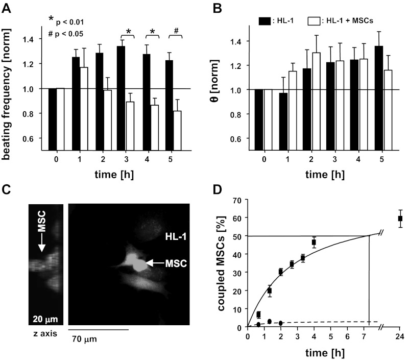
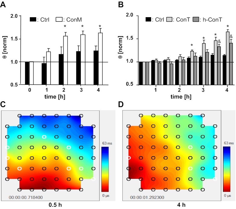
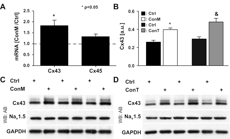
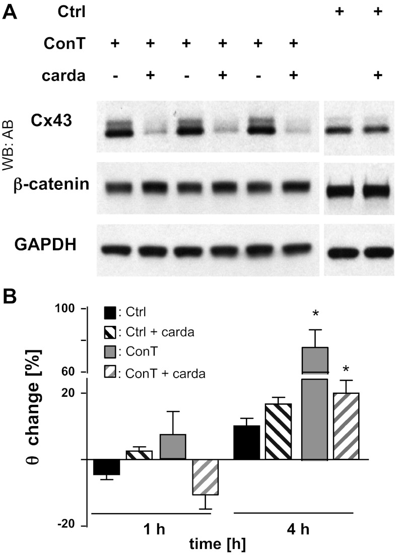
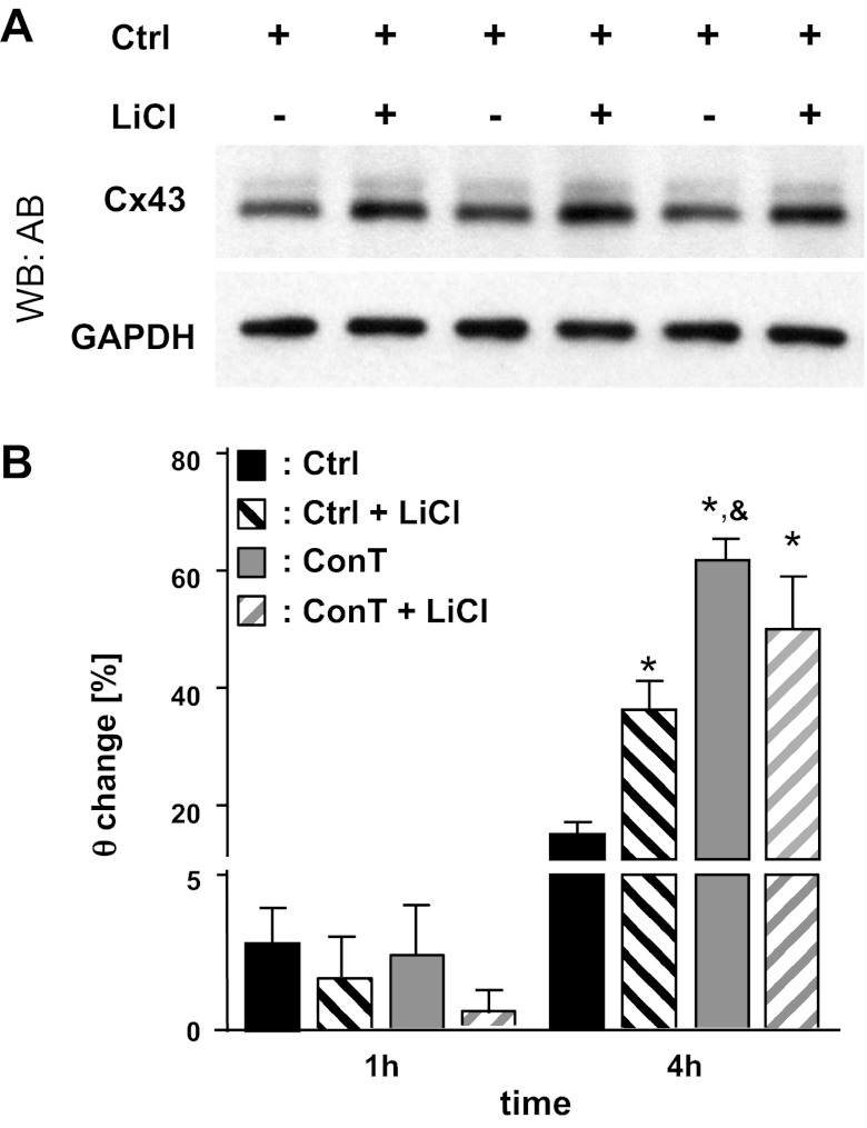
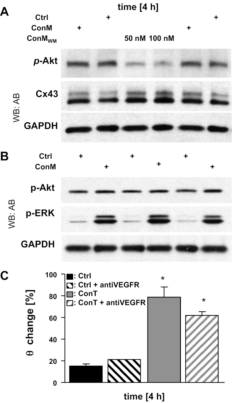
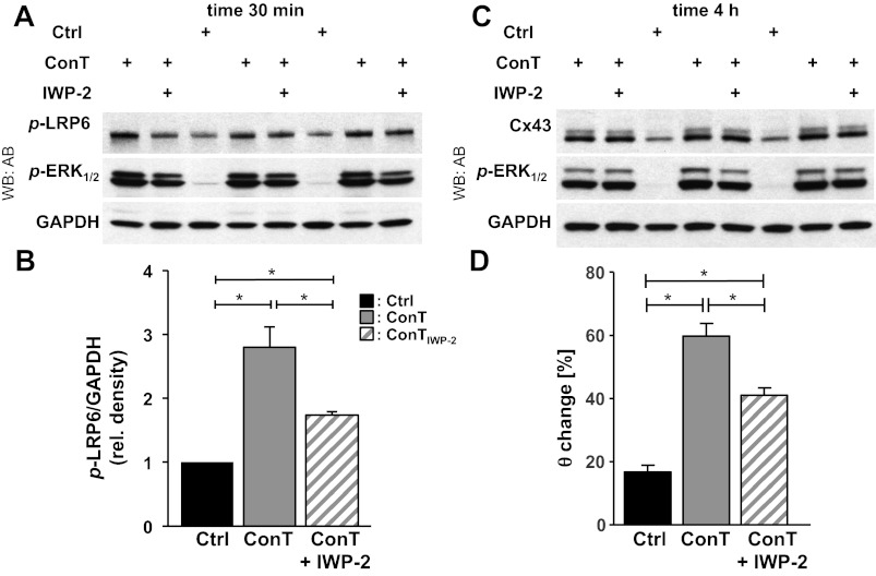
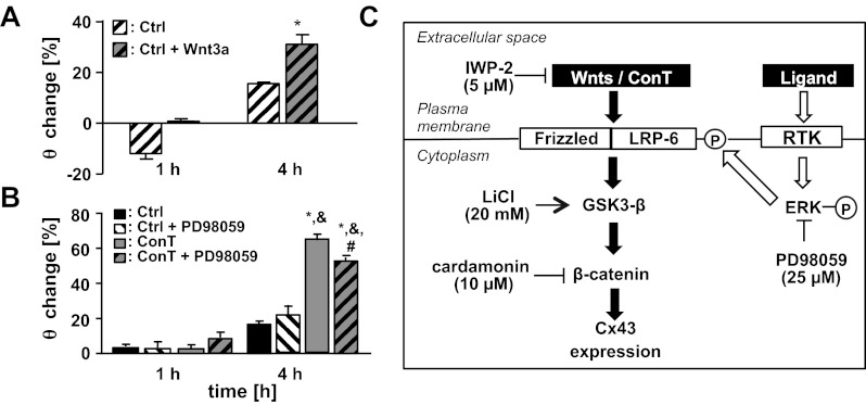
Mesenchymal stem cells (MSCs) were shown to improve cell survival and alleviate cardiac arrhythmias when transplanted into cardiac tissue; however, little is known about the mechanism by which MSCs modify the electrophysiological properties of cardiac tissue. We aimed to distinguish the influence of cell-cell coupling between myocytes and MSCs from that of MSC-derived paracrine factors on the spontaneous activity and conduction velocity (θ) of multicellular cardiomyocyte preparations. HL-1 cells were plated on microelectrode arrays and their spontaneous activity and θ was determined from field potential recordings. In heterocellular cultures of MSCs and HL-1 cells the beating frequency was attenuated (t(0h): 2.26 ± 0.18 Hz; t(4h): 1.98 ± 0.26 Hz; P < 0.01) concomitant to the intercellular coupling between MSCs and cardiomyocytes. In HL-1 monolayers supplemented with MSC conditioned media (ConM) or tyrode (ConT) θ significantly increased in a time-dependent manner (ConT: t(0h): 2.4 cm/s ± 0.2; t(4h): 3.1 ± 0.4 cm/s), whereas the beating frequency remained constant. Connexin (Cx)43 mRNA and protein expression levels also increased after ConM or ConT treatment over the same time period. Enhanced low-density lipoprotein receptor-related protein 6 (LRP6) phosphorylation after ConT treatment implicates the Wnt signaling pathway. Suppression of Wnt secretion from MSCs (IWP-2; 5 μmol/l) reduced the efficacy of ConT to induce phospho-LRP6 and to increase θ. Inhibition of β-catenin (cardamonin; 10 μmol/l) or GSK3-α/β (LiCl; 5 mmol/l) also suppressed changes in θ, further supporting the hypothesis that MSC-mediated Cx43 upregulation occurs in part through secreted Wnt ligands and activation of the canonical Wnt signaling pathway.
Figures
Similar articles
-
Excitation-contraction coupling in ventricular myocytes is enhanced by paracrine signaling from mesenchymal stem cells.J Mol Cell Cardiol. 2012 Jun;52(6):1249-56. doi: 10.1016/j.yjmcc.2012.03.008. Epub 2012 Mar 23. J Mol Cell Cardiol. 2012. PMID: 22465692 Free PMC article.
-
Lithium stimulates human bone marrow derived mesenchymal stem cell proliferation through GSK-3β-dependent β-catenin/Wnt pathway activation.FEBS J. 2014 Dec;281(23):5371-89. doi: 10.1111/febs.13081. Epub 2014 Oct 27. FEBS J. 2014. PMID: 25265417
-
Activation of canonical wnt pathway promotes differentiation of mouse bone marrow-derived MSCs into type II alveolar epithelial cells, confers resistance to oxidative stress, and promotes their migration to injured lung tissue in vitro.J Cell Physiol. 2013 Jun;228(6):1270-83. doi: 10.1002/jcp.24282. J Cell Physiol. 2013. PMID: 23154940
-
Intracellular Signals Activated by Canonical Wnt Ligands Independent of GSK3 Inhibition and β-Catenin Stabilization.Cells. 2019 Sep 25;8(10):1148. doi: 10.3390/cells8101148. Cells. 2019. PMID: 31557964 Free PMC article. Review.
-
Paracrine Mechanisms Involved in Mesenchymal Stem Cell Differentiation into Cardiomyocytes.Curr Stem Cell Res Ther. 2019;14(1):9-13. doi: 10.2174/1574888X13666180821160421. Curr Stem Cell Res Ther. 2019. PMID: 30152289 Review.
Cited by
-
Evidence for Transfer of Membranes from Mesenchymal Stem Cells to HL-1 Cardiac Cells.Stem Cells Int. 2014;2014:653734. doi: 10.1155/2014/653734. Epub 2014 Sep 9. Stem Cells Int. 2014. PMID: 25295065 Free PMC article.
-
Slow conduction in mixed cultured strands of primary ventricular cells and stem cell-derived cardiomyocytes.Front Cell Dev Biol. 2015 Sep 24;3:58. doi: 10.3389/fcell.2015.00058. eCollection 2015. Front Cell Dev Biol. 2015. PMID: 26442264 Free PMC article.
-
Restoring heart function and electrical integrity: closing the circuit.NPJ Regen Med. 2017 Apr 7;2:9. doi: 10.1038/s41536-017-0015-2. eCollection 2017. NPJ Regen Med. 2017. PMID: 29302345 Free PMC article. Review.
-
Connexins modulate autophagosome biogenesis.Nat Cell Biol. 2014 May;16(5):401-14. doi: 10.1038/ncb2934. Epub 2014 Apr 6. Nat Cell Biol. 2014. PMID: 24705551 Free PMC article.
-
The role of P21-activated kinase (Pak1) in sinus node function.J Mol Cell Cardiol. 2023 Jun;179:90-101. doi: 10.1016/j.yjmcc.2023.04.004. Epub 2023 Apr 20. J Mol Cell Cardiol. 2023. PMID: 37086972 Free PMC article.
References
-
- Aberg ND, Blomstrand F, Aberg MA, Bjorklund U, Carlsson B, Carlsson-Skwirut C, Bang P, Ronnback L, Eriksson PS. Insulin-like growth factor-I increases astrocyte intercellular gap junctional communication and connexin43 expression in vitro. J Neurosci Res 74: 12–22, 2003 - PubMed
-
- Ai X, Pogwizd SM. Connexin 43 downregulation and dephosphorylation in nonischemic heart failure is associated with enhanced colocalized protein phosphatase type 2A. Circ Res 96: 54–63, 2005 - PubMed
-
- Angoulvant D, Ivanes F, Ferrera R, Matthews PG, Nataf S, Ovize M. Mesenchymal stem cell conditioned media attenuates in vitro and ex vivo myocardial reperfusion injury. J Heart Lung Transplant 30: 95–102, 2011 - PubMed
Publication types
MeSH terms
Substances
Grants and funding
LinkOut - more resources
Full Text Sources
Other Literature Sources