. 2013 Feb 15;304(4):H579-88.
doi: 10.1152/ajpheart.00754.2012. Epub 2012 Dec 15.

Functional differences between junctional and extrajunctional adrenergic receptor activation in mammalian ventricle

Affiliations

Functional differences between junctional and extrajunctional adrenergic receptor activation in mammalian ventricle

Olujimi A Ajijola et al. Am J Physiol Heart Circ Physiol. .

Abstract

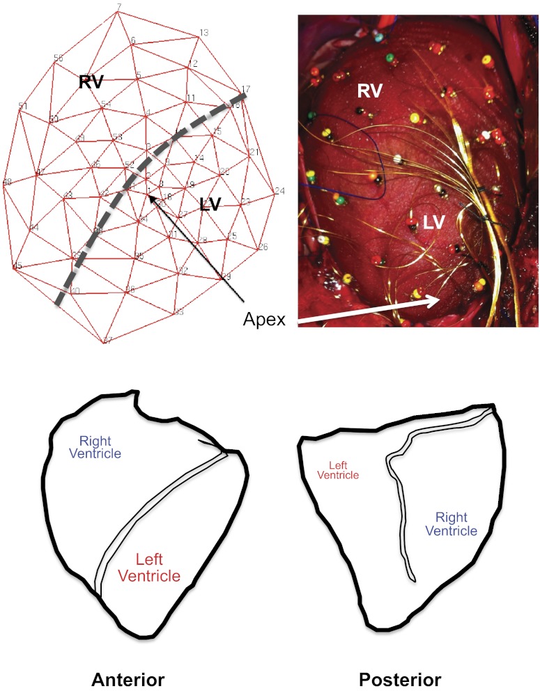
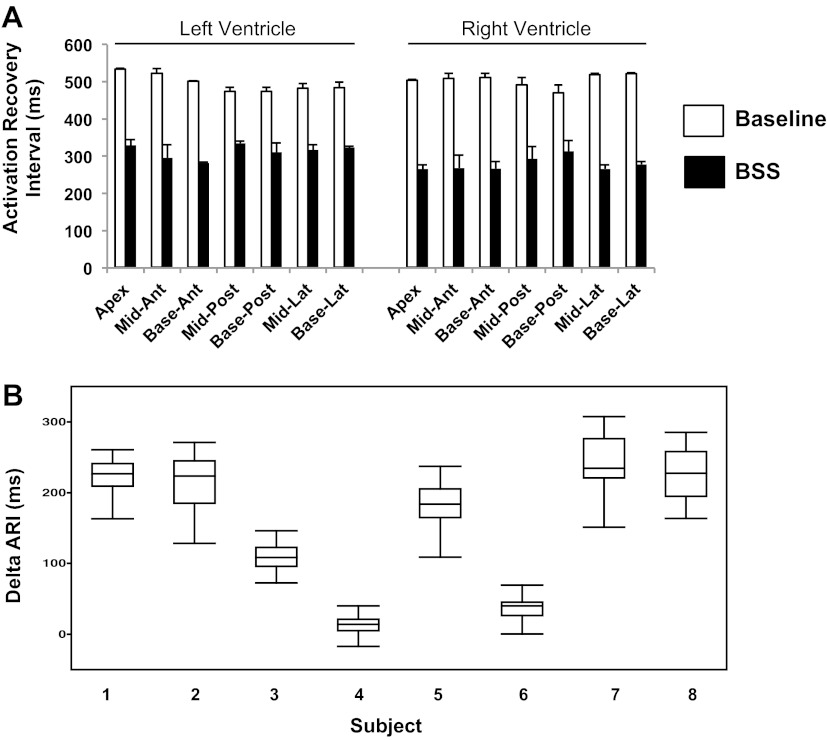
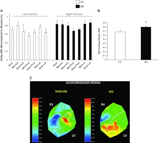
Increased cardiac sympathetic activation worsens dispersion of repolarization and is proarrhythmic. The functional differences between intrinsic nerve stimulation and adrenergic receptor activation remain incompletely understood. This study was undertaken to determine the functional differences between efferent cardiac sympathetic nerve stimulation and direct adrenergic receptor activation in porcine ventricles. Female Yorkshire pigs (n = 13) underwent surgical exposure of the heart and stellate ganglia. A 56-electrode sock was placed over the ventricles to record epicardial electrograms. Animals underwent bilateral sympathetic stimulation (BSS) (n = 8) or norepinephrine (NE) administration (n = 5). Activation recovery intervals (ARIs) were measured at each electrode before and during BSS or NE. The degree of ARI shortening during BSS or NE administration was used as a measure of functional nerve or adrenergic receptor density. During BSS, ARI shortening was nonuniform across the epicardium (F value 9.62, P = 0.003), with ARI shortening greatest in the mid-basal lateral right ventricle and least in the midposterior left ventricle (LV) (mean normalized values: 0.9 ± 0.08 vs. 0.56 ± 0.08; P = 0.03). NE administration resulted in greater ARI shortening in the LV apex than basal segments [0.91 ± 0.04 vs. 0.63 ± 0.05 (averaged basal segments); P = 0.003]. Dispersion of ARIs increased in 50% and 60% of the subjects undergoing BSS and NE, respectively, but decreased in the others. There is nonuniform response to cardiac sympathetic activation of both porcine ventricles, which is not fully explained by adrenergic receptor density. Different pools of adrenergic receptors may mediate the cardiac electrophysiological effects of efferent sympathetic nerve activity and circulating catecholamines.


Figures

Fig. 1.
Fig. 1.
Configuration of the 56-electrode sock and porcine ventricular anatomy. A: schematic representation of the sock electrode including relative positions of the right ventricle (RV) and left ventricle (LV) (left). After placement on porcine ventricles, the actual 56-electrode sock is shown, with relative position of the RV and LV (right). B: relationship of the porcine RV and LV in the anterior and posterior orientation (left and right, respectively).
Fig. 2.
Fig. 2.
Hemodynamic response to bilateral sympathetic stimulation. A: hemodynamic tracings from a left ventricular conductance catheter showing the response to bilateral sympathetic stimulation (BSS). Graphical representations of the change in heart rate (HR; B), systolic blood pressure (SBP) and left ventricular end-systolic pressure (LV-ESP) (C), and inotropy (dP/dtmax) and lusitropy (dP/dtmin) (D) are shown. *P < 0.05, ***P < 0.001, ****P < 0.0001 (n = 5 for SBP and n = 8 for HR, LV-ESP, dP/dtmax, and dP/dtmin).
Fig. 3.
Fig. 3.
BSS and norepinephrine (NE) administration alter epicardial electrograms. A representative sample of 5 electrograms distributed over the porcine LV and RV is shown. These were recorded at baseline and after 5 min of BSS (A) and baseline and at peak NE effect (B). Both tracings from BSS and NE administration show shortening of the R-R and QT intervals, and altered repolarization demonstrated by T wave changes.
Fig. 4.
Fig. 4.
BSS induces global activation recovery interval shortening. A: representative example of activation recovery intervals (ARIs) from 7 LV and 7 RV regions, at baseline and after BSS. Means ± SD are shown. B: change in ARI (ΔARI) across all 8 subjects studied. Subjects 4 and 6 showed ARI prolongation in some leads. Ant, anterior; lat, lateral; post, posterior.
Fig. 5.
Fig. 5.
Functional density of cardiac sympathetic nerves. A: degree of ARI shortening induced by BSS in all 14 regions across the LV and RV is shown, normalized to the maximum. ARI shortening was uneven across both ventricles (F statistic, 9.62; P = 0.003; n = 8). B: ARI shortening was greater in the RV than the LV (P = 0.016, n = 8). C: representative polar maps showing ARI distribution at baseline and after BSS. The mid- to basal lateral RV showed the shortest ARI at baseline, but shortened to a greater degree when compared with other regions. Ant, anterior; lat, lateral; post, posterior. *P < 0.05.
Fig. 6.
Fig. 6.
NE administration induces global activation recovery interval shortening. A: representative example of ARIs from 7 LV and 7 RV regions, at baseline and after NE administration. Means ± SD are shown. B: change in ARI (ΔARI) across all 5 subjects that underwent NE administration is shown. One pig (subject 3) showed ARI prolongation in some leads. Ant, anterior; lat, lateral; post, posterior.
Fig. 7.
Fig. 7.
Functional density of cardiac β-adrenergic receptors. A: degree of ARI shortening induced by NE administration in all 14 regions across the LV and RV is shown, normalized to the maximum. ARI shortening was uneven across the left ventricle. ARI shortening in the LV apex was significantly greater than in all basal segments. B: there was no difference between ARI shortening on the LV or RV (P = 0.73, n = 5). C: representative polar maps showing ARI distribution at baseline and at peak NE administration. The LV apex showed the greater ARI shortening (blue region) than all LV basal segments. The RV did not show a similar pattern of apex-base gradient in ARI. Ant, anterior; lat, lateral; post, posterior.
Fig. 8.
Fig. 8.
Apico-basal density of cardiac sympathetic nerves and cardiac β-adrenergic receptors. ARI change normalized to the maximum is displayed for the apical and basal regions of the LV and RV for A. Bilateral sympathetic stimulation showed no significant apical basal shortening for either the LV or RV (n = 8). B: NE administration resulted in greater ARI shortening in the LV apex than the basal segments of the LV. No apico-basal differences were seen for the RV (n = 5). Ant, anterior; lat, lateral; post, posterior.
Fig. 9.
Fig. 9.
Dispersion of ARIs during bilateral sympathetic stimulation and NE administration. The dispersion of ARIs (determined as variance of ARI values) at baseline and during BSS (A) or NE administration (B) is shown for all subjects studied. For both conditions, some subjects showed an increase in ARI dispersion, whereas others showed decrease.

References

    1. Amerini S, Rubino A, Filippi S, Ledda F, Mantelli L. Modulation by adrenergic transmitters of the efferent function of capsaicin-sensitive nerves in cardiac tissue. Neuropeptides 20: 225–232, 1991 - PubMed
    1. Angelakos ET. Regional distribution of catecholamines in the dog heart. Circ Res 16: 39–44, 1965 - PubMed
    1. Angelakos ET, Fuxe K, Torchiana ML. Chemical and histochemical evaluation of the distribution of catecholamines in the rabbit and guinea pig hearts. Acta physiologica Scandinavica 59: 184–192, 1963 - PubMed
    1. Basu S, Sinha SK, Shao Q, Ganguly PK, Dhalla NS. Neuropeptide Y modulation of sympathetic activity in myocardial infarction. Journal of the American College of Cardiology 27: 1796–1803, 1996 - PubMed
    1. Bristow MR, Ginsburg R, Umans V, Fowler M, Minobe W, Rasmussen R, Zera P, Menlove R, Shah P, Jamieson S. Beta 1- and beta 2-adrenergic-receptor subpopulations in nonfailing and failing human ventricular myocardium: coupling of both receptor subtypes to muscle contraction and selective beta 1-receptor down-regulation in heart failure. Circ Res 59: 297–309, 1986 - PubMed

Publication types

MeSH terms

LinkOut - more resources