. 2012 Dec;16(12):3116-22.
doi: 10.1111/j.1582-4934.2012.01582.x.

Telocytes in the human kidney cortex

Affiliations

Telocytes in the human kidney cortex

Guisheng Qi et al. J Cell Mol Med. 2012 Dec.

Abstract

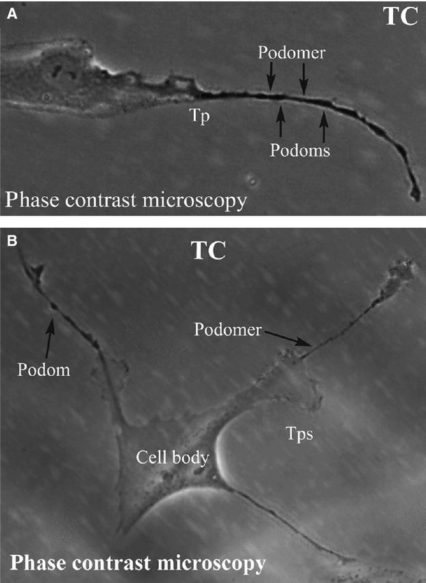
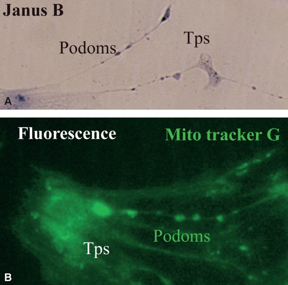
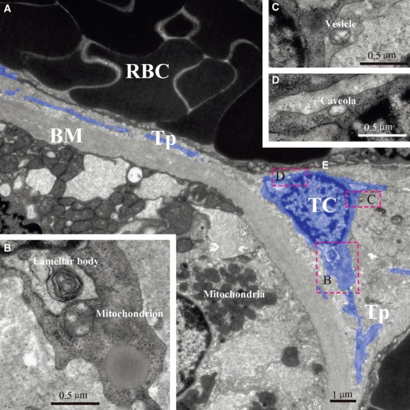
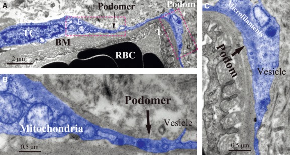
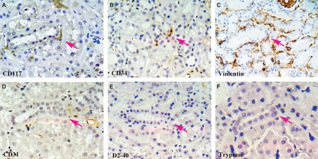
Renal interstitial cells play an important role in the physiology and pathology of the kidneys. As a novel type of interstitial cell, telocytes (TCs) have been described in various tissues and organs, including the heart, lung, skeletal muscle, urinary tract, etc. (www.telocytes.com). However, it is not known if TCs are present in the kidney interstitium. We demonstrated the presence of TCs in human kidney cortex interstitium using primary cell culture, transmission electron microscopy (TEM) and in situ immunohistochemistry (IHC). Renal TCs were positive for CD34, CD117 and vimentin. They were localized in the kidney cortex interstitial compartment, partially covering the tubules and vascular walls. Morphologically, renal TCs resemble TCs described in other organs, with very long telopodes (Tps) composed of thin segments (podomers) and dilated segments (podoms). However, their possible roles (beyond intercellular signalling) as well as their specific phenotype in the kidney remain to be established.


Figures

Fig 1
Fig 1
(A) and (B) Phase contrast microscopy of kidney TCs in primary culture. Note the typically very long Tps (over 50 μm). Also, the specific structure of Tps is obvious: alternation of dilations (podoms) with thin segments (podomers). Direct magnification: 400×. TC: Telocyte; Tp: Telopode.
Fig 2
Fig 2
(A) and (B) Kidney TCs in primary culture. (A) Vital staining for mitochondria with Janus Green B; note the podoms coloured blue-brown because of the presence of mitochondria. (B) Fluorescence microscopy using Mito Tracker Green  as molecular probe for the presence of mitochondria; note the moniliform aspect of Tps as a result of accumulation of mitochondria in podoms. TC: Telocyte; Tp: Telopode; Mito Tracker G: Mito Tracker Green.
Fig 3
Fig 3
Immunofluorescence labelling for CD117, CD34 and vimentin of cultured cells. (A) FITC labelling for CD117 (green). (B) FITC labelling for CD34 (yellow). (C) TRITC labelling for vimentin (red). DAPI staining for nuclei (blue) in (A), (B), and (C). FITC: fluorescein isothiocyanate; TRITC: tetraethyl rhodamine isothiocyanate; DAPI: 4′, 6-diamidino-2-phenylindole.
Fig 4
Fig 4
TEM image of kidney telocyte. There is a typical telocyte between a tubule and a vessel (A). Mitochondrion and lamellar body are found near nuclear (B). Shed vesicle and caveola could also be found after magnification (C) and (D). TC: telocyte; Tp: telopode; RBC: red blood cell; E: endothelium; BM: basement membrane.
Fig 5
Fig 5
TEM image of kidney telocyte. A typical telocyte with Tps is around a blood capillary located in kidney interstitium (A). Note Tps which prolong along the basement membrane of vessel. Details of its podomer and podom are presented (B) and (C). Shed vesicle is next to podomer and obvious mitochondria are next to nuclear (B). In podom, there are microfilaments and vesicle (C). TC: telocyte; RBC: red blood cell; E: endothelium; BM: basement membrane.
Fig 6
Fig 6
In situ IHC of CD117, CD34, vimentin, CD31, D2–40 and Tryptase on serial slides. One cell with positive expression of CD117 can be seen in the kidney interstitium (A; magenta arrow). It has three long and thin Tps. Positive expressions of CD34 and vimentin can be observed in the same area of serial slides (B) and (C). Staining results of CD31, D2–40 and tryptase are presented (D)–(F) on serial slides. Their negative expressions in same area help us exclude mast cell staining of CD117 and endothelial cell staining of CD34. Magnification: 400×.

References

    1. Popescu LM, Faussone-Pellegrini MS. TELOCYTES - a case of serendipity: the winding way from Interstitial Cells of Cajal (ICC), via Interstitial Cajal-Like Cells (ICLC) to TELOCYTES. J Cell Mol Med. 2010;14:729–40. - PMC - PubMed
    1. Kostin S. Myocardial telocytes: a specific new cellular entity. J Cell Mol Med. 2010;14:1917–21. - PMC - PubMed
    1. Popescu LM, Manole CG, Gherghiceanu M, et al. Telocytes in human epicardium. J Cell Mol Med. 2010;14:2085–93. - PMC - PubMed
    1. Gherghiceanu M, Manole CG, Popescu LM. Telocytes in endocardium: electron microscope evidence. J Cell Mol Med. 2010;14:2330–4. - PMC - PubMed
    1. Suciu L, Nicolescu MI, Popescu LM. Cardiac telocytes: serial dynamic images in cell culture. J Cell Mol Med. 2010;14:2687–92. - PMC - PubMed

Publication types