. 2013 Feb;33(2):305-10.
doi: 10.1161/ATVBAHA.112.300485. Epub 2012 Dec 13.

Reduced mural cell coverage and impaired vessel integrity after angiogenic stimulation in the Alk1-deficient brain

Affiliations

Reduced mural cell coverage and impaired vessel integrity after angiogenic stimulation in the Alk1-deficient brain

Wanqiu Chen et al. Arterioscler Thromb Vasc Biol. 2013 Feb.

Abstract

Objective: Vessels in brain arteriovenous malformations are prone to rupture. The underlying pathogenesis is not clear. Hereditary hemorrhagic telangiectasia type 2 patients with activin receptor-like kinase 1 (Alk1) mutation have a higher incidence of brain arteriovenous malformation than the general population. We tested the hypothesis that vascular endothelial growth factor impairs vascular integrity in the Alk1-deficient brain through reduction of mural cell coverage.

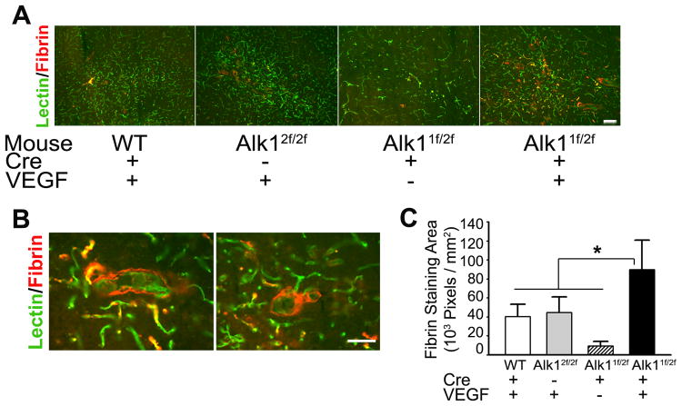
Methods and results: Adult Alk1(1f/2f) mice (loxP sites flanking exons 4-6) and wild-type mice were injected with 2×10(7) PFU adenovious-cre recombinase and 2×10(9) genome copies of adeno-associated virus-vascular endothelial growth factor to induce focal homozygous Alk1 deletion (in Alk1(1f/2f) mice) and angiogenesis. Brain vessels were analyzed 8 weeks later. Compared with wild-type mice, the Alk1-deficient brain had more fibrin (99±30×10(3) pixels/mm(2) versus 40±13×10(3); P=0.001), iron deposition (508±506 pixels/mm(2) versus 6±49; P=0.04), and Iba1(+) microglia/macrophage infiltration (888±420 Iba1(+) cells/mm(2) versus 240±104 Iba1(+); P=0.001) after vascular endothelial growth factor stimulation. In the angiogenic foci, the Alk1-deficient brain had more α-smooth muscle actin negative vessels (52±9% versus 12±7%, P<0.001), fewer vascular-associated pericytes (503±179/mm(2) versus 931±115, P<0.001), and reduced platelet-derived growth factor receptor-β expression.

Conclusions: Reduction of mural cell coverage in response to vascular endothelial growth factor stimulation is a potential mechanism for the impairment of vessel wall integrity in hereditary hemorrhagic telangiectasia type 2-associated brain arteriovenous malformation.


Figures

Figure 1
Figure 1
The dysplastic vessels in the angiogenic foci of Alk1-deficient brain have increased permeability than vessels in the angiogenic foci of the normal brain. (A) Representative images show vessels (lectin, green) and fibrin (red) in the viral-injected region. (B) High magnification images show the location of fibrin. The fibrin lines outside the dysplastic vessel wall in the Alk1-deficient brain. (C) Bar graph shows the quantification of fibrin. In the viral-injected region, there were significantly more fibrin deposition in the VEGF-treated Alk1-deficient brain than VEGF-treated WT (*p= 0.001) and Alk12f/2f (*p= 0.003) groups, and Alk1-deficient brain without VEGF treatment (*p<0.001). Scale bars: 50 μm.
Figure 2
Figure 2
RBCs and iron deposition were detected around the dysplastic vessels. (A) Representative images of Prussian blue staining show the iron deposition around dysplastic vessels in the angiogenic foci of Alk1-deficient brain. (B) Bar graph shows the quantification of Prussian blue-positive area. The VEGF-stimulated Alk1-deficient group had significantly increased Prussian blue-positive area than VEGF-treated WT (*p=0.04) and Alk12f/2f (*p=0.03) group, and untreated Alk1-deficient brain (*p=0.02). (C) RBCs were found in extravascular space (black arrow). Scale bar: 50 μm. The insert shows a macrophage with three red blood cells in its cytoplasm. Scale bar in the insert: 10 μm. The brain was perfused with PBS before collection. (D) Representative images of hematoxylin and eosin (H&E) and Prussian blue stained Alk1-deficient mouse and human bAVM sections. Hemosiderin and Prussian blue-positive staining were detected around dysplastic vessels in the angiogenic foci of Alk1-deficient brain and in the human bAVM. Scale bars: 50 μm.
Figure 3
Figure 3
Prussian blue staining positively correlated with macrophage/microglia infiltration. (A) Representative images of Iba1 antibody staining (red). Scale bar: 50 μm. (B) Bar graph shows quantification of Iba1+ cells. The VEGF-stimulated Alk1-deficient group had more Iba1+ cells than VEGF-treated WT (*p=0.001) and Alk12f/2f (*p=0.015) groups, and untreated Alk1-deficient-group (*p=0.002). (C) Graph shows Prussian blue-positive area positively correlated with the number of Iba1+ cells (R2=0.2, *p<0.05).
Figure 4
Figure 4
The dysplastic vessels in the angiogenic foci of Alk1-deficient brain had less coverage of α-SMA+ cells. (A) Representative images of lectin (green) and anti-αSMA (red) stained sections. In the angiogenic foci of Alk1-deficient brain, some vessels larger than 15 μm had no α-SMA+ cells (white arrows). Scale bar: 50 μm. (B) Bar graph shows the percentage of α-SMA- vessels (>15 μm). Significantly more α-SMA- vessels presented in the VEGF-stimulated Alk1-deficient brain than that in WT (*p<0.001), Alk12f/2f (*p<0.001) and Alk1-deficient-only (*p<0.001) groups. (C) Prussian Blue stains were detected around α-SMA- vessels. The left panel shows Prussian blue-positive staining around the α-SMA- vessels, such as Vessels a and b (black arrows). Scale bars: 50 μm. The right panel shows high magnification images of Vessel a from the left panel. H&E (top) and Prussian blue (bottom) stained sections. Brown colored cells around vessels are hemosiderin positive macrophages. Scale bars: 30 μm. (D) Pie graphs show significantly more PB+ vessels are α-SMA- vessels (*p<0.001). PB: Prussian blue.
Figure 5
Figure 5
The dysplastic vessels in the angiogenic foci of Alk1-deficient brain have fewer pericytes on their walls. (A) Representative images show vessels (lectin, green) and pericytes (upper panel: NG2, red; lower panel: vascular associated ZIC1+ cells, red) in the vector injection sites. * in upper panel and arrows in lower panel indicate dysplastic vessels. Scale bars: 50 μm. (B) High magnification images of pericytes. The upper panel shows vascular (green) associated NG2+ pericyte (arrows, red). The lower panel shows the ZIC1+ pericytes (arrows, red) co-localized with nuclei (DAPI, blue). Scale bars: 20 μm. (C) Bar graph shows the quantification of pericytes located on the vessel wall. (D) Graph shows the number of vessel-associated pericyte inversely correlating with the degree of fibrin deposition (R2 =0.45).

References

    1. Attia W, Tada T, Hongo K, Nagashima H, Takemae T, Tanaka Y, Kobayashi S. Microvascular pathological features of immediate perinidal parenchyma in cerebral arteriovenous malformations: giant bed capillaries. J Neurosurg. 2003;98:823–827. - PubMed
    1. Sato S, Kodama N, Sasaki T, Matsumoto M, Ishikawa T. Perinidal dilated capillary networks in cerebral arteriovenous malformations. Neurosurgery. 2004;54:163–168. discussion 168-170. - PubMed
    1. Kim H, Marchuk DA, Pawlikowska L, Chen Y, Su H, Yang GY, Young WL. Genetic considerations relevant to intracranial hemorrhage and brain arteriovenous malformations. Acta Neurochir Suppl. 2008;105:199–206. - PMC - PubMed
    1. Walker EJ, Su H, Shen F, Choi EJ, Oh SP, Chen G, Lawton MT, Kim H, Chen Y, Chen W, Young WL. Arteriovenous malformation in the adult mouse brain resembling the human disease. Ann Neurol. 2011;69:954–962. - PMC - PubMed
    1. Park SO, Wankhede M, Lee YJ, Choi EJ, Fliess N, Choe SW, Oh SH, Walter G, Raizada MK, Sorg BS, Oh SP. Real-time imaging of de novo arteriovenous malformation in a mouse model of hereditary hemorrhagic telangiectasia. J Clin Invest. 2009;119:3487–3496. - PMC - PubMed

Publication types

MeSH terms

Supplementary concepts