. 2013 Jan;123(1):443-54.
doi: 10.1172/JCI65831. Epub 2012 Dec 17.

Annexin A1, formyl peptide receptor, and NOX1 orchestrate epithelial repair

Affiliations

Annexin A1, formyl peptide receptor, and NOX1 orchestrate epithelial repair

Giovanna Leoni et al. J Clin Invest. 2013 Jan.

Abstract

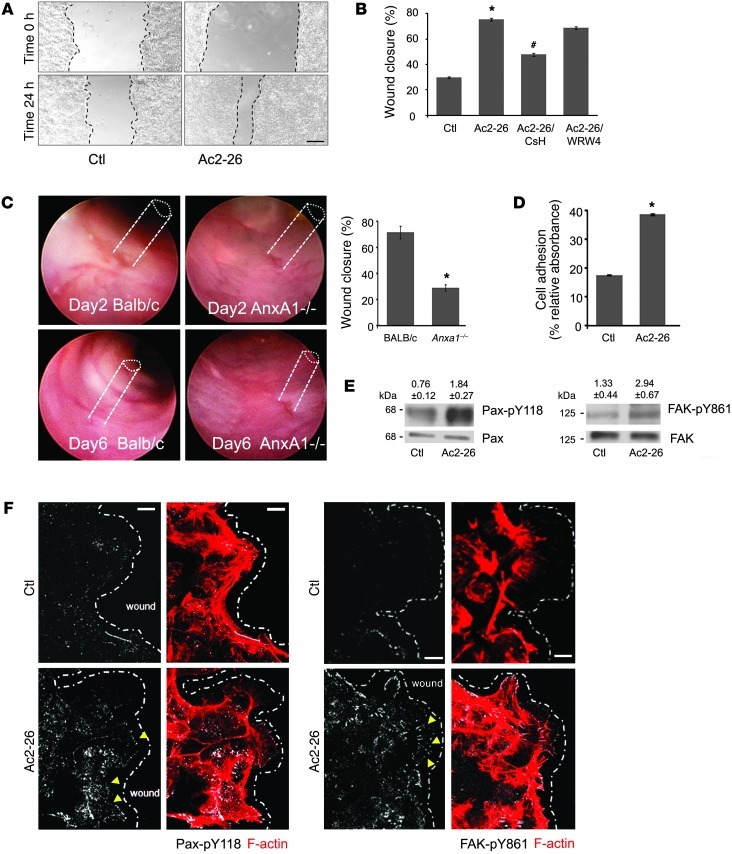
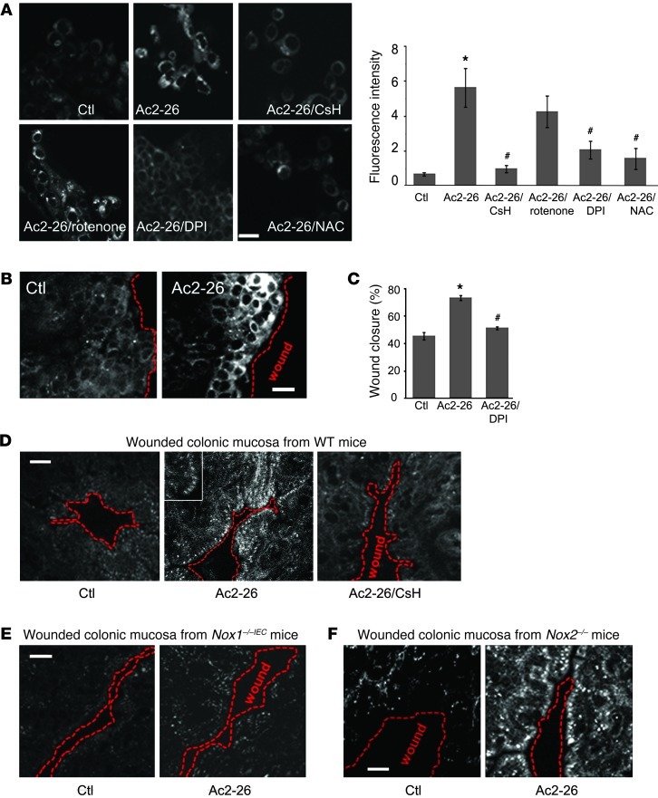
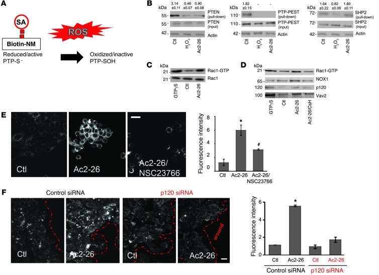
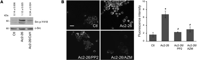
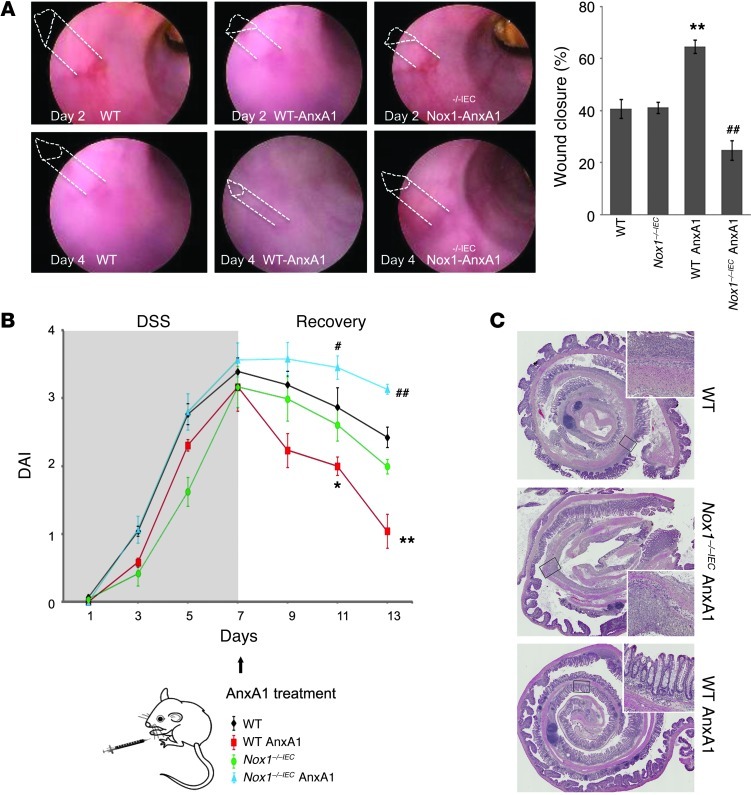
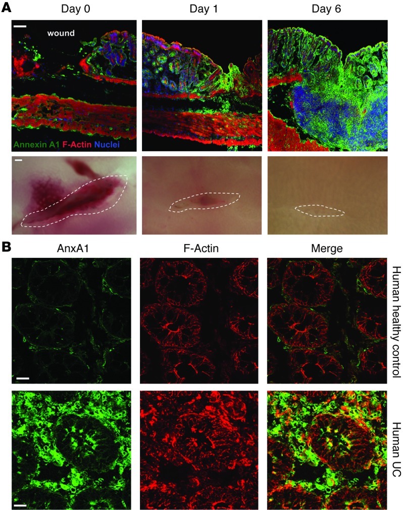
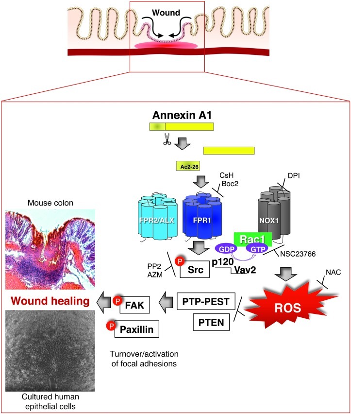
N-formyl peptide receptors (FPRs) are critical regulators of host defense in phagocytes and are also expressed in epithelia. FPR signaling and function have been extensively studied in phagocytes, yet their functional biology in epithelia is poorly understood. We describe a novel intestinal epithelial FPR signaling pathway that is activated by an endogenous FPR ligand, annexin A1 (ANXA1), and its cleavage product Ac2-26, which mediate activation of ROS by an epithelial NADPH oxidase, NOX1. We show that epithelial cell migration was regulated by this signaling cascade through oxidative inactivation of the regulatory phosphatases PTEN and PTP-PEST, with consequent activation of focal adhesion kinase (FAK) and paxillin. In vivo studies using intestinal epithelial specific Nox1(-/-IEC) and AnxA1(-/-) mice demonstrated defects in intestinal mucosal wound repair, while systemic administration of ANXA1 promoted wound recovery in a NOX1-dependent fashion. Additionally, increased ANXA1 expression was observed in the intestinal epithelium and infiltrating leukocytes in the mucosa of ulcerative colitis patients compared with normal intestinal mucosa. Our findings delineate a novel epithelial FPR1/NOX1-dependent redox signaling pathway that promotes mucosal wound repair.


Figures

Figure 1
Figure 1. ANXA1 regulates wound healing via FPR1 signaling in IECs and stimulates phosphorylation of focal adhesion proteins (FAK and Pax).
(A) SK-CO15 monolayers were subjected to scratch wound assay in the presence of Ac2-26 peptide (3 μM). Wound widths were determined at 0 and 24 hours. Photomicrograph shows representative results for control (Ctl) and Ac2-26–treated cells. Scale bar: 200 μm. (B) SK-CO15 cells were also incubated with Ac2-26 and CsH (1 μM) or WRW4 (10 μM). The ANXA1 cleavage product Ac2-26 alone significantly enhanced wound closure (*P < 0.0001). The increase in wound closure was inhibited in the presence of CsH (#P < 0.0001) but not WRW4. The experiment was repeated 3 times, and results of 1 representative experiment done with 5 parallel samples are shown. (C) Endoscopic images of colonic mucosal wounds in mice (AnxA1–/– and control BALB/c) at days 2 and 4 after injury. Quantification of wound repair is shown in the graph (mean ± SEM, *P = 0.023, n = 11 mice/group). (D) SK-CO15 cells were plated on ECL gel, and non-adherent cells were removed by washing at 1 hour after plating. Adhesion increased within 15 minutes of Ac2-26 exposure compared with non-stimulated cells (*P < 0.0001). (E) Immunoblot of SK-CO15 cells revealed a significant increase in Pax phosphorylation (Y118, 2.4-fold) and FAK phosphorylation (Y861, 2.2-fold) in cells stimulated with Ac2-26 for 15 minutes compared with unstimulated cells (0.1% DMSO). Normalized signal intensity is indicated above the blots. (F) Laser confocal micrographs of Pax p-Y118 (white) or FAK p-Y861 (white) and F-actin (red) in migrating SK-CO15 cells with or without Ac2-26 (3 μM) for 15 minutes. Photomicrographs are representative of 3 independent experiments done in triplicate. Scale bars: 10 μm.
Figure 2
Figure 2. Ac2-26 peptide induces rapid ROS generation in IECs via FPR1 and NOX1.
(A) SK-CO15 cells were incubated with Ac2-26 peptide for 15 minutes, and ROS generation was detected by confocal microscopy using the fluorescent hydro-Cy3 dye. Ac2-26 treatment increased fluorescence intensity, which was inhibited by CsH, DPI, and NAC, but not by rotenone. Summarized data for hydro-Cy3 fluorescence intensity are presented in the graph (mean ± SEM,*P < 0.05 vs. control, #P < 0.05 vs. Ac2-26, n = 3). (B) ROS generation in scratch-wounded SK-CO15 monolayer was increased in cells adjoining the wound. Confocal micrographs are representative of 3 independent experiments (C) SK-CO15 monolayers were subjected to scratch wound assay in the presence of Ac2-26 (3 μM) with or without DPI. Wound widths were determined at 0 and 24 hours (mean ± SEM, *P < 0.05 vs. Ctl, #P < 0.05 vs. Ac2-26). (D) Whole mount preparations of colon taken from WT (D), Nox1–/–IEC (E), and Nox2–/– (F) mice injected with hydro-Cy3 (i.p., 30 minutes) were mechanically wounded ex vivo. Inset in D highlights ROS generation at the leading edge of migrating cells. Mucosa was luminally treated for 15 minutes with vehicle (Ctl) or Ac2-26 with or without preincubation of the tissue samples with CsH. Confocal micrographs are representative of 3 independent experiments (5 mice/group). Scale bars: 40 μm.
Figure 3
Figure 3. Ac2-26 oxidizes PTP-PEST and PTEN and activates Rac1-GTPases.
(A) ROS oxidize thiols in redox-sensitive phosphatases (PTPs). Maleimide groups react efficiently and specifically with reduced sulfhydryls (R-S) of PTPs to form stable thioester bonds but do not react and bind R-SOH oxidized sulfenic acid (thioester) forms. (B) Cell were treated with vehicle, H2O2 (15 μM), or Ac2-26 (3 μM) for 15 minutes and subjected to labeling with biotin-NM. Biotinylated proteins were precipitated with SA beads and subjected to immunoblot analysis using PTEN, PTP-PEST, and SHP2 antibodies. β-Actin was used as a loading control. Immunoblots presented are representative of 3 independent experiments. Normalized signal intensity is indicated above the blots. (C) SK-CO15 cells were incubated for 30 minutes at room temperature with 100 μM GTPγS, vehicle, or Ac2-26 peptide for 15 minutes. Active Rac1 (Rac1-GTP) was pulled down using agarose bead–conjugated GST-PAK-RBD. Total (Rac1) and active Rac1 (Rac1-GTP) were analyzed by immunoblot. Immunoblots are representative of 3 independent experiments. (D) Active Rac1 pull-down samples were subjected to immunoblot analysis with antibodies against Vav2, p120, and NOX1. Immunoblots are representative of 3 independent experiments. (E) ROS generation was analyzed in SK-CO15 cells, treated with hydro-Cy3 dye, by confocal microscopy. Cells were incubated with vehicle, Ac2-26 (3 μM), or Ac2-26 plus NSC23766. Micrographs are representative of 3 independent experiments. Scale bar: 20 μm. (F) SK-CO15 cells were transfected with siRNA targeting p120 catenin. Three days after siRNA transfection, cells were treated with Ac2-26 for 15 minutes, and ROS generation was detected. Confocal micrographs are representative of 4 independent experiments. Scale bar: 20 μm. Summarized data for hydro-Cy3 fluorescence intensity are presented in the graphs (mean ± SEM,*P < 0.05 vs control, #P < 0.05 vs. Ac2-26, n = 3).
Figure 4
Figure 4. Ac2-26 peptide activates Src kinase.
(A) Immunoblots of phospho-Src (Src p-Y418) and total Src (c-Src) expression in SK-CO15 cells stimulated with control (0.1% DMSO) and Ac2-26 (3 μM) for 15 minutes. Immunoblots are representative of 3 independent experiments. Normalized signal intensity is indicated above the blots. (B) Confocal images of ROS-sensitive hydro-Cy3 in SK-CO15 cells treated with vehicle, Ac2-26 (3 μM), or Ac2-26 (3 μM) after preincubation with PP2 and AZM. Confocal micrographs are representative of 4 independent experiments. Scale bar: 20 μm. Summarized data for hydro-Cy3 fluorescence intensity are presented in the graph (mean ± SEM,*P < 0.05 vs. control, #P < 0.05 vs. Ac2-26 n = 4).
Figure 5
Figure 5. ANXA1 accelerates in vivo intestinal mucosal wound healing and recovery via NOX1.
(A) Endoscopic images of healing colonic mucosal wounds 2 or 4 days after biopsy-induced injury in WT and Nox1–/–IEC mice treated with i.p. injections of PBS or ANXA1 (5 μg, twice/day). Quantification of wound repair is shown in the graph. ANXA1 administration promoted recovery of wounds in WT mice, but in the absence of NOX1 resulted in a significant delay in wound healing. (B) Clinical disease activity index (DAI) of mice subjected to DSS colitis for 7 days, followed by recovery from colitis for 6 days. ANXA1 was administered i.p. (5 μg, twice/day) during recovery. Significantly decreased DAI was observed in WT mice treated with ANXA1 (red line) compared with control mice treated with PBS (black line). However, ANXA1 failed to enhance recovery in Nox1–/–IEC mice (blue line). (C) Representative photomicrographs of H&E-stained histological sections. Boxed areas are magnified in insets. The total magnification of the photomicrographs is ×2 and in the insets it is ×40. Data in all graphs are presented as mean ± SEM; *P < 0.05 and **P < 0.001, WT ANXA1 vs. WT PBS, n = 12 mice/group; #P < 0.05 and ##P < 0.01, Nox1–/–IEC ANXA1 vs. Nox1–/–IEC PBS, n = 12 mice/group.
Figure 6
Figure 6. Mucosal ANXA1 expression is increased in injured murine and human colonic tissue.
(A) Representative images of colonic mucosa from WT mice 0, 1, and 6 days after biopsy-induced injury. Frozen sections were stained with antibodies against ANXA1 (green), F-actin (phalloidin, red), and nuclei (TO-PRO-3, blue). Scale bar: 100 μm. The bottom row shows photomicrographs of colonic mucosa wounds on days 0, 1, and 6; n = 5 mice/group. (B) Sections of human colonic tissue from healthy control and UC patients were stained with ANXA1 (green) and F-actin (red). Scale bars: 20 μm.
Figure 7
Figure 7. Model for the molecular mechanism of ANXA1-induced NOX1-dependent redox signaling during epithelial wound repair.
Ac2-26 binds to FPR1, which induces Src activation (Src p-Y418), resulting in the formation of a complex comprising active Rac1, p120 catenin, Vav2, and NOX1. ROS generated by NOX1 induces oxidative inactivation of PTEN and PTP-PEST, leading to increased phosphorylation of the focal adhesion proteins FAK and Pax, thereby promoting epithelial cell movement and wound repair.

Similar articles

Cited by

References

    1. Lotz MM, Nusrat A, Madara JL, Ezzell R, Wewer UM, Mercurio AM. Intestinal epithelial restitution. Involvement of specific laminin isoforms and integrin laminin receptors in wound closure of a transformed model epithelium. Am J Pathol. 1997;150(2):747–760. - PMC - PubMed
    1. Perretti M, et al. Endogenous lipid- and peptide-derived anti-inflammatory pathways generated with glucocorticoid and aspirin treatment activate the lipoxin A4 receptor. Nat Med. 2002;8(11):1296–1302. doi: 10.1038/nm786. - DOI - PMC - PubMed
    1. Serhan CN. The resolution of inflammation: the devil in the flask and in the details. FASEB J. 2011;25(5):1441–1448. doi: 10.1096/fj.11-0502ufm. - DOI - PMC - PubMed
    1. Serhan CN. Systems approach to inflammation resolution: identification of novel anti-inflammatory and pro-resolving mediators. J Thromb Haemost. 2009;7(suppl 1):44–48. doi: 10.1111/j.1538-7836.2009.03396.x. - DOI - PubMed
    1. Serhan CN, et al. Resolution of inflammation: state of the art, definitions and terms. FASEB J. 2007;21(2):325–332. doi: 10.1096/fj.06-7227rev. - DOI - PMC - PubMed

Publication types

MeSH terms