. 2013 Jan;31(1):71-5.
doi: 10.1038/nbt.2459. Epub 2012 Dec 16.

Combinatorial antigen recognition with balanced signaling promotes selective tumor eradication by engineered T cells

Affiliations

Combinatorial antigen recognition with balanced signaling promotes selective tumor eradication by engineered T cells

Christopher C Kloss et al. Nat Biotechnol. 2013 Jan.

Abstract

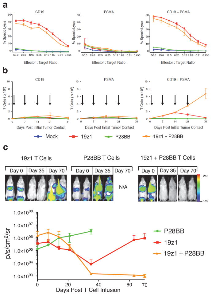
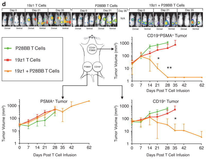
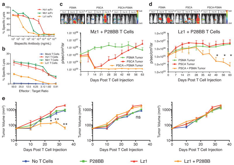
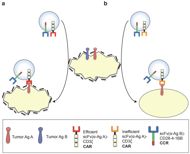
Current T-cell engineering approaches redirect patient T cells to tumors by transducing them with antigen-specific T-cell receptors (TCRs) or chimeric antigen receptors (CARs) that target a single antigen. However, few truly tumor-specific antigens have been identified, and healthy tissues that express the targeted antigen may undergo T cell-mediated damage. Here we present a strategy to render T cells specific for a tumor in the absence of a truly tumor-restricted antigen. T cells are transduced with both a CAR that provides suboptimal activation upon binding of one antigen and a chimeric costimulatory receptor (CCR) that recognizes a second antigen. Using the prostate tumor antigens PSMA and PSCA, we show that co-transduced T cells destroy tumors that express both antigens but do not affect tumors expressing either antigen alone. This 'tumor-sensing' strategy may help broaden the applicability and avoid some of the side effects of targeted T-cell therapies.


Figures

Figure 1
Figure 1. Dual chimeric receptor-mediated activation and costimulation of human T cells facilitates robust cytotoxicity, proliferation, and tumor eradication
(a,b) T cells were mock transduced or were transduced with retroviruses encoding a CD19-specific CAR (19z1) and/or a PSMA-specific CCR (P28BB). (a) T cells were incubated at indicated effector:target ratios with 51Cr-loaded target cells expressing CD19 and/or PSMA, and target cell lysis (chromium release) was measured. Plots represent at least 4 experiments, with error bars representing standard deviation of the mean of 3 replicates. (b) T cells were co-cultured with PC3 tumor cell lines expressing CD19 and/or PSMA (arrows indicate re-stimulation of T cells using freshly irradiated tumor cells). T cell numbers were measured at indicated time intervals. Plots represent at least 4 experiments with error bars representing standard deviation of the mean of 3 replicates. (c) T cells (1.0 × 10⁶) described in (a,b) were injected intravenously into NSG mice bearing green fluorescent protein/firefly-luciferase fusion protein (GFP/Luc) expressing CD19+PSMA+ PC3 human prostate tumors. Tumor burden was measured weekly by bioilluminescent imaging. Top, images of two representative mice from each group at each time point, with the pixel intensity represented in color. Bottom, average tumor burden as quantified by luminescence of the tumors using units of photons per second per square centimeter per steradian (p/sec/cm2/sr); error bars represent standard deviation from the mean. Values were generated from 6 mice per group. (d) 1 × 10⁶ CD19+PSMA, 1 × 10⁶ CD19PSMA+ and 1 × 10⁶ CD19+PSMA+ PC3 cells were injected subcutaneously into the left flank, right flank and back, respectively, of NSG mice. T cells expressing 19z1 and/or P28BB were infused intravenously 7 days later. Top, representative images of 2 mice per time point per group as analyzed by bioluminescent imaging. Graphs, tumors were quantitatively measured using calipers and tumor volumes were plotted versus time for each tumor. Error bars represent standard deviation from the mean of 6 mice. Statistical significance was determined using two-tailed unpaired t tests to compare values obtained from 19z1 T cells and 19z1 + P28BB T cells and p values are represented as * for <0.05 or ** for <0.01.
Figure 2
Figure 2. Reducing CAR mediated activation signals restricts T cell activity to tumors expressing both antigens upon combinatorial antigen recognition
(a) Three different single chain variable fragments (scFvs) specific for PSCA were constructed and assembled into bispecific antibodies in which the other scFv binds to CD3. The bispecific antibodies were added in the indicated amounts to cocultures of T cells and 51Cr-labeled PSCA+ PC3 tumor cells (ratio of 20:1). Specific lysis was measured by chromium release. Graph is a representative of n > 4 experiments. (b) The anti-PSCA scFvs described in (a) were used to generate CARs. T cells were mock transduced or were transduced with retroviruses encoding these CARs, and were indubated with 51Cr-labeled PSCA+ PC3 tumor cells at indicated effector:target ratios. Specific lysis was measured by chromium release. Graph is a representative of n > 3 experiments. (c,d) Green fluorescent protein/firefly-luciferase fusion protein (GFP/Luc) expressing PC3 tumor cells expressing PSMA and/or PSCA were injected intravenously into the tail-vein of NSG mice. Fourteen days later, 1.0 × 10⁶ Mz1 +P28BB (c) or Lz1 + P28BB (d) T cells were infused intravenously. Top, bioluminescent imaging of two representative mice from each group at each time point. Graphs, the average tumor burden was quantified by luminescence of the tumors using units of photons per second per square centimeter per steradian (p/sec/cm2/sr) and plotted with error bars representing standard deviation from the mean of values from 5 mice per group. Two mice that received PSMA+ tumors (green line) died after day 49 and therefore the mean value for luminescence was averaged from 3 values for days 56 and 63. (e) 0.5 × 10⁶ CD19+PSMA, 0.5 × 10⁶ CD19PSMA+ and 0.5 × 10⁶ CD19+PSMA+ PC3 cells were injected subcutaneously into the left flank, right flank and back, respectively, of NSG mice. Where indicated 1.0 × 10⁶ CAR+ T cells transduced with Lz1 and/or P28BB were infused intravenously 7 days later. Tumor volume was measured at indicated time points and plotted with error bars representing standard deviation from the mean of values from 5 mice per each group except for 10 mice for Lz1 + P28BB group). Statistical significance was determined using two-tailed unpaired t tests to compare values obtained from Lz1 T cells and Lz1 + P28BB T cells and p values are represented as * for <0.05 or ** for <0.01.
Figure 3
Figure 3. Concept of combinatorial antigen recognition and balanced signaling for selective tumor eradication
(a) T cells expressing an efficient CAR (specific for antigen A) and a CCR (specific for antigen B), become potently activated and costimulated by A+B+ target cells, which are eliminated. If these T cells recirculate (indicated by arrows) and encounter A+B target cells, they can eliminate these as well without requiring further costimulation, resulting in an “on-target off-tumor” effect. (b) In the tumor-sensing approach, T cells expressing a CCR and an inefficient activation receptor (e.g. low affinity TCR or suboptimal CAR) are sufficiently activated by A+B+ but not A+B target cells.

Comment in

References

    1. Sadelain M, Riviere I, Brentjens R. Targeting tumours with genetically enhanced T lymphocytes. Nature reviews. Cancer. 2003;3:35–45. - PubMed
    1. Ho WY, Blattman JN, Dossett ML, Yee C, Greenberg PD. Adoptive immunotherapy: engineering T cell responses as biologic weapons for tumor mass destruction. Cancer cell. 2003;3:431–437. - PubMed
    1. Sadelain M, Brentjens R, Riviere I. The promise and potential pitfalls of chimeric antigen receptors. Curr Opin Immunol. 2009;21:215–223. - PMC - PubMed
    1. Robbins PF, et al. Tumor regression in patients with metastatic synovial cell sarcoma and melanoma using genetically engineered lymphocytes reactive with NY-ESO-1. Journal of clinical oncology : official journal of the American Society of Clinical Oncology. 2011;29:917–924. - PMC - PubMed
    1. Kalos M, et al. T cells with chimeric antigen receptors have potent antitumor effects and can establish memory in patients with advanced leukemia. Sci Transl Med. 2011;3:95ra73. - PMC - PubMed

Publication types