

Distinct neural mechanisms of distractor suppression in the frontal and parietal lobe
- PMID: 23242309
- PMCID: PMC4207121
- DOI: 10.1038/nn.3282
Distinct neural mechanisms of distractor suppression in the frontal and parietal lobe
Abstract
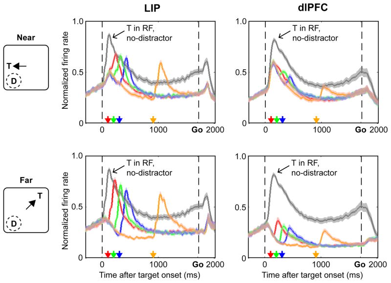
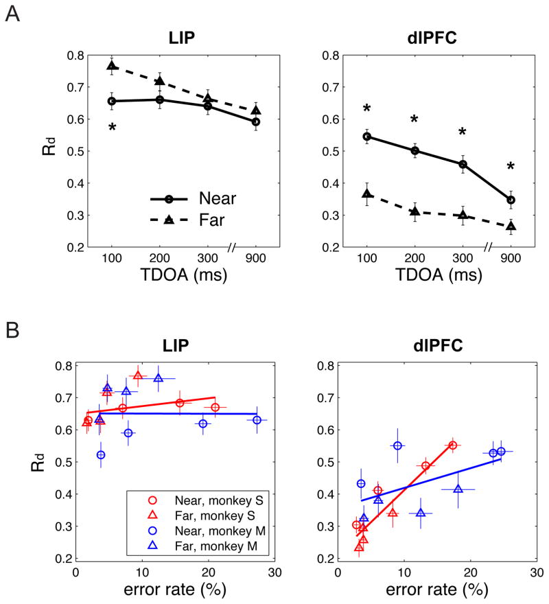
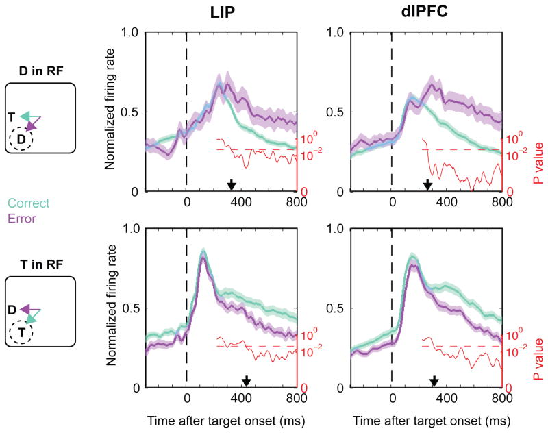
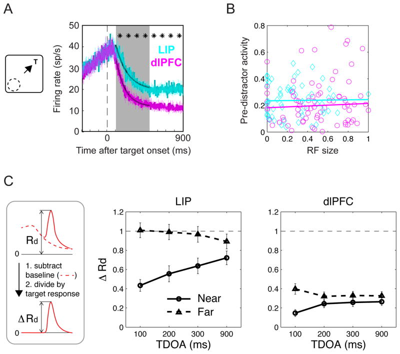
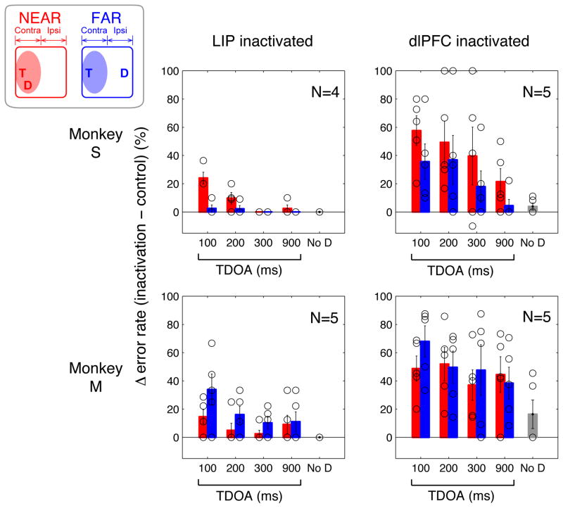
The posterior parietal cortex and the prefrontal cortex are associated with eye movements and visual attention, but their specific contributions are poorly understood. We compared the dorsolateral prefrontal cortex (dlPFC) and the lateral intraparietal area (LIP) in monkeys using a memory saccade task in which a salient distractor flashed at a variable timing and location during the memory delay. We found that the two areas had similar responses to target selection, but made distinct contributions to distractor suppression. Distractor responses were more strongly suppressed and more closely correlated with performance in the dlPFC relative to LIP. Moreover, reversible inactivation of the dlPFC produced much larger increases in distractibility than inactivation of LIP. These findings suggest that LIP and dlPFC mediate different aspects of selective attention. Although both areas can contribute to the perceptual selection of salient information, the dlPFC has a decisive influence on whether and how attended stimulus is linked with actions.
Conflict of interest statement
The authors declare that they have no conflict of interest.
Figures

Comment in
-
Parietal and prefrontal neurons driven to distraction.Nat Neurosci. 2013 Jan;16(1):8-9. doi: 10.1038/nn.3291. Nat Neurosci. 2013. PMID: 23257928 No abstract available.
Similar articles
-
Response of neurons in the lateral intraparietal area to a distractor flashed during the delay period of a memory-guided saccade.J Neurophysiol. 2000 Jul;84(1):301-10. doi: 10.1152/jn.2000.84.1.301. J Neurophysiol. 2000. PMID: 10899205
-
Neural correlates of attention and distractibility in the lateral intraparietal area.J Neurophysiol. 2006 Mar;95(3):1696-717. doi: 10.1152/jn.00848.2005. Epub 2005 Dec 7. J Neurophysiol. 2006. PMID: 16339000 Free PMC article.
-
Motor intention activity in the macaque's lateral intraparietal area. I. Dissociation of motor plan from sensory memory.J Neurophysiol. 1996 Sep;76(3):1439-56. doi: 10.1152/jn.1996.76.3.1439. J Neurophysiol. 1996. PMID: 8890265
-
Saccades, salience and attention: the role of the lateral intraparietal area in visual behavior.Prog Brain Res. 2006;155:157-75. doi: 10.1016/S0079-6123(06)55010-1. Prog Brain Res. 2006. PMID: 17027387 Free PMC article. Review.
-
The relationship between spatial attention and saccades in the frontoparietal network of the monkey.Eur J Neurosci. 2011 Jun;33(11):1973-81. doi: 10.1111/j.1460-9568.2011.07710.x. Eur J Neurosci. 2011. PMID: 21645093 Review.
Cited by
-
Transcranial magnetic stimulation over the right dorsolateral prefrontal cortex modulates visuospatial distractor suppression.Eur J Neurosci. 2021 May;53(10):3394-3403. doi: 10.1111/ejn.15164. Epub 2021 Mar 17. Eur J Neurosci. 2021. PMID: 33650122 Free PMC article.
-
Coding of latent variables in sensory, parietal, and frontal cortices during closed-loop virtual navigation.Elife. 2022 Oct 25;11:e80280. doi: 10.7554/eLife.80280. Elife. 2022. PMID: 36282071 Free PMC article.
-
Stable Working Memory and Perceptual Representations in Macaque Lateral Prefrontal Cortex during Naturalistic Vision.J Neurosci. 2022 Nov 2;42(44):8328-8342. doi: 10.1523/JNEUROSCI.0597-22.2022. Epub 2022 Oct 4. J Neurosci. 2022. PMID: 36195438 Free PMC article.
-
High-definition transcranial direct current stimulation dissociates fronto-visual theta lateralization during visual selective attention.J Physiol. 2020 Mar;598(5):987-998. doi: 10.1113/JP278788. Epub 2020 Feb 3. J Physiol. 2020. PMID: 31840247 Free PMC article. Clinical Trial.
-
Priming by motivationally salient distractors produces hemispheric asymmetries in visual processing.Psychol Res. 2019 Nov;83(8):1798-1807. doi: 10.1007/s00426-018-1028-1. Epub 2018 May 24. Psychol Res. 2019. PMID: 29797045
References
-
- Badre D, Wagner AD. Left ventrolateral prefrontal cortex and the cognitive control of memory. Neuropsychologia. 2007;45:2883–2901. - PubMed
-
- Corbetta M, Shulman GL. Control of goal-directed and stimulus-driven attention in the brain. Nat Rev Neurosci. 2002;3:201–215. - PubMed
-
- Egeth HE, Yantis S. Visual attention: control, representation, and time course. Annu Rev Psychol. 1997;48:269–297. - PubMed
-
- Klingberg T. Development of a superior frontal-intraparietal network for visuo-spatial working memory. Neuropsychologia. 2006;44:2171–2177. - PubMed
Publication types
MeSH terms
Substances
Grants and funding
LinkOut - more resources
Full Text Sources
