

Review
doi: 10.3390/v4123754.
Use of the Syrian hamster as a new model of ebola virus disease and other viral hemorrhagic fevers
Affiliations
- PMID: 23242370
- PMCID: PMC3528289
- DOI: 10.3390/v4123754
Item in Clipboard
Review
Use of the Syrian hamster as a new model of ebola virus disease and other viral hemorrhagic fevers
Viruses.
.
Abstract
Historically, mice and guinea pigs have been the rodent models of choice for therapeutic and prophylactic countermeasure testing against Ebola virus disease (EVD). Recently, hamsters have emerged as a novel animal model for the in vivo study of EVD. In this review, we discuss the history of the hamster as a research laboratory animal, as well as current benefits and challenges of this model. Availability of immunological reagents is addressed. Salient features of EVD in hamsters, including relevant pathology and coagulation parameters, are compared directly with the mouse, guinea pig and nonhuman primate models.
Figures
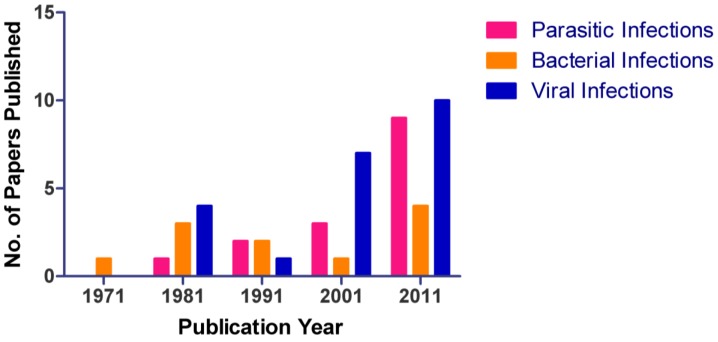
Number of publications utilizing hamsters between 1971 through 2011. An increase in use of hamsters as an animal model for parasitic, bacterial, and viral diseases is noted in the literature. During the 10-year period of 2001-2011, the largest increase in publications was observed, the majority of which were in the virology sector.
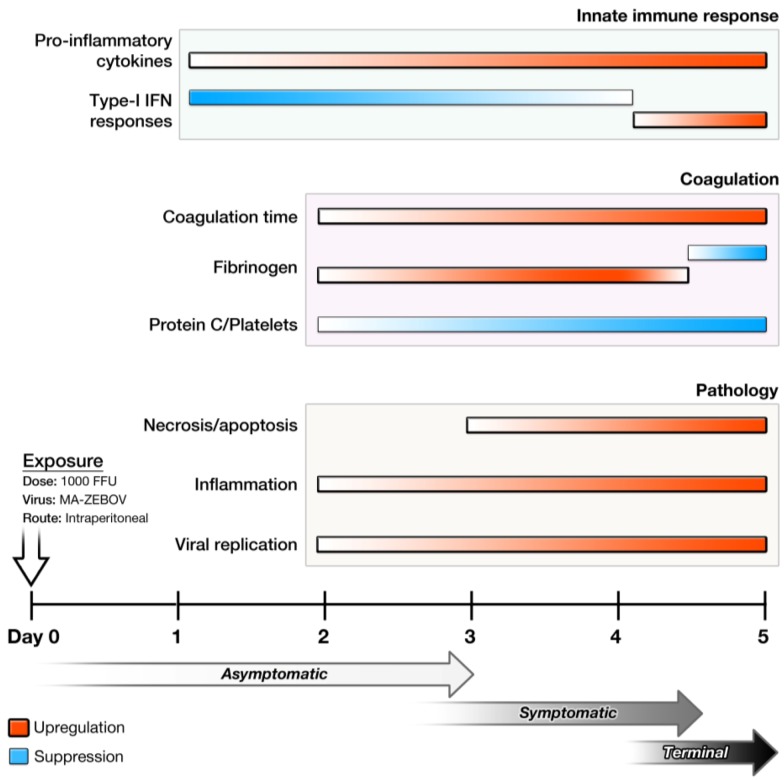
Temporal progression of disease in the Syrian hamster model of Ebola virus disease. Following exposure to 1000 focus-forming units of MA-EBOV IP, hamsters begin showing signs of illness around day 3. Changes in the innate immune response, coagulation parameters, and pathology are observed as early as days 1 and 2.
Comparison of pathology in mouse, guinea pig, hamster, and nonhuman primate. A Balb/c mouse and a Syrian hamster were infected IP with MA-EBOV; a Hartley strain of guinea pig was infected with GPA-EBOV; and a macaque was infected with wild-type EBOV. (A-D) Pathological changes in liver of different animal models. (A) Mouse: Multifocal, random hepatocellular degeneration and necrosis (10x and 40x inset). (B) Guinea pig: Diffuse, random hepatocellular degeneration and necrosis. Inflammatory cells are nearly absent (10x and 40x inset). (C–D) Hamsters: Liver. (C) Diffuse, midzonal hepatocellular degeneration, necrosis, and congestion. Inflammatory cells are nearly absent (10x). Solid arrow: prominent intracytoplasmic filovirus inclusion bodies in hepatocytes (40x). (D) Diffuse, random hepatocellular degeneration and necrosis (10x). Solid star: fibrin deposition (40x inset). (E-L) Pathological changes in spleen of different animal models. (E and I) Mouse: White and red pulp. White pulp (E); diffuse lymphoid necrosis and loss (10x and 40x inset). Red pulp (I); mild to moderate acute splenitis and small amounts of fibrin (solid star) (40x). (F and J) Guinea pig: White and red pulp. White pulp (F); multifocal lymphoid necrosis (10x and 40x inset). Red pulp (J); multifocal, mild to moderate acute splenitis with necrosis. Solid star: small amounts of fibrin at marginal zone (40x). (G and K) Hamster: White and red pulp. White pulp (G); diffuse lymphoid necrosis (10x and 40x inset). Red pulp (K); mild to moderate acute splenitis with monocytic degeneration and necrosis (40x). (H and L) NHP: White and red pulp. White pulp (H); diffuse lymphoid necrosis (10x and 20x inset). Red pulp (L); diffuse, moderate acute splenitis (40x). Solid star: fibrin.
References
-
- Schnittler H.J., Feldmann H. Viral hemorrhagic fever--a vascular disease? Thromb. Haemostasis. 2003;89:967–972. - PubMed
-
- Zaki S.R., Goldsmith C.S. Pathologic features of filovirus infections in humans. Curr. Top. Microbiol. Immunol. 1999;235:97–116. - PubMed
-
- Bwaka M.A., Bonnet M.J., Calain P., Colebunders R., De Roo A., Guimard Y., Katwiki K.R., Kibadi K., Kipasa M.A., Kuvula K.J., et al. Ebola hemorrhagic fever in Kikwit, Democratic Republic of the Congo: clinical observations in 103 patients. J. Infect. Dis. 1999;179:S1–S7. doi: 10.1086/314568. - DOI - PubMed
-
- Formenty P., Hatz C., Le Guenno B., Stoll A., Rogenmoser P., Widmer A. Human infection due to Ebola virus, subtype Cote d'Ivoire: Clinical and biologic presentation. J. Infect. Dis. 1999;179:S48–S53. - PubMed
-
- Kuhn J.H. Filoviruses. A compendium of 40 years of epidemiological, clinical, and laboratory studies. Arch. Virol. Suppl. 2008;20:13–360. - PubMed
Publication types
MeSH terms
Grants and funding
LinkOut - more resources
Full Text Sources
Other Literature Sources
Medical
