. 2012 Dec 14;4(12):3785-803.
doi: 10.3390/v4123785.

Diversity in glycosaminoglycan binding amongst hMPV G protein lineages

Affiliations

Diversity in glycosaminoglycan binding amongst hMPV G protein lineages

Penelope Adamson et al. Viruses. .

Abstract

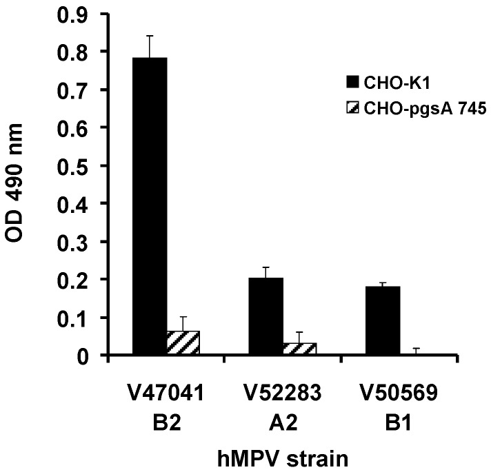
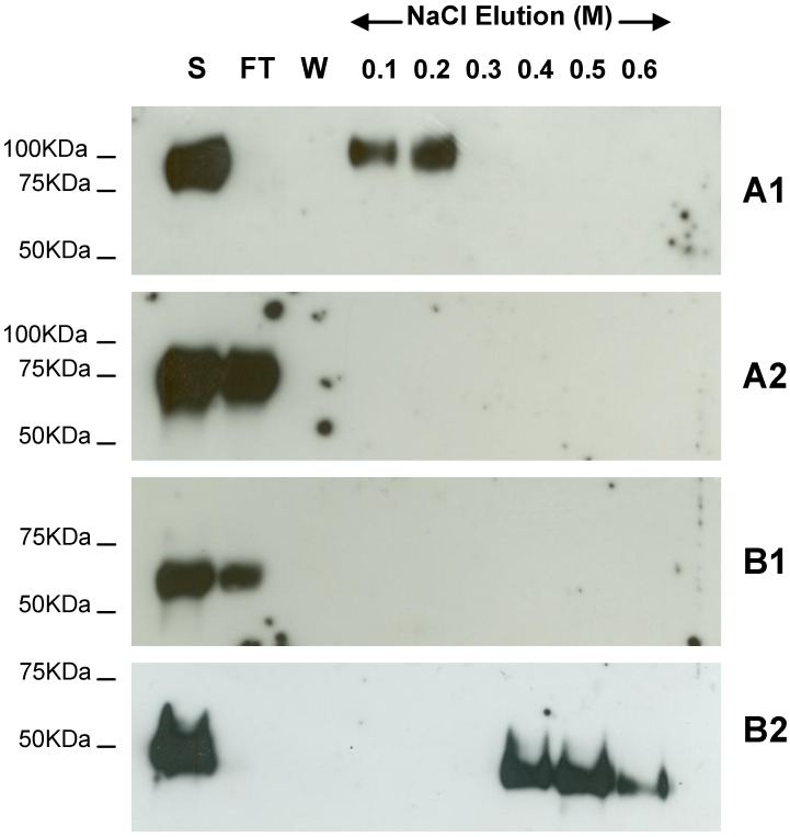
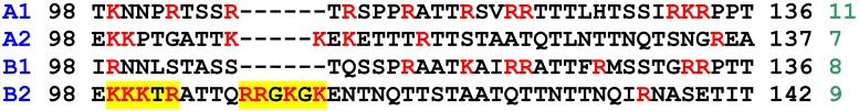
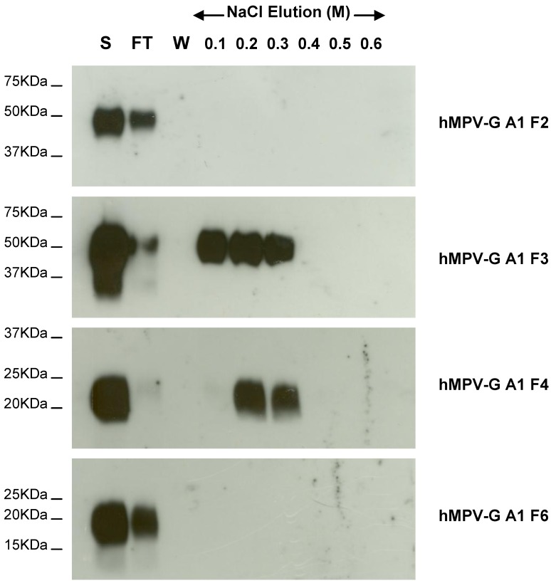
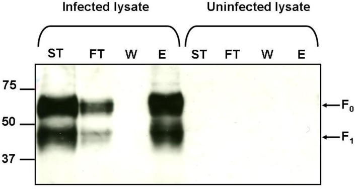
We have previously shown that hMPV G protein (B2 lineage) interacts with cellular glycosaminoglycans (GAGs). In this study we examined subtypes A1, A2 and B1 for this interaction. GAG-dependent infectivity of available hMPV strains was demonstrated using GAG-deficient cells and heparin competition. We expressed the G protein ectodomains from all strains and analysed these by heparin affinity chromatography. In contrast to the B2 lineage, neither the A2 or B1 G proteins bound to heparin. Sequence analysis of these strains indicated that although there was some homology with the B2 heparin-binding domains, there were less positively charged residues, providing a likely explanation for the lack of binding. Although sequence analysis did not demonstrate well defined positively charged domains in G protein of the A1 strain, this protein was able to bind heparin, albeit with a lower affinity than G protein of the B2 strain. These results indicate diversity in GAG interactions between G proteins of different lineages and suggest that the GAG-dependency of all strains may be mediated by interaction with an alternative surface protein, most probably the conserved fusion (F) protein. Analysis of both native and recombinant F protein confirmed that F protein binds heparin, supporting this conclusion.


Figures

Figure 1
Figure 1
Primary isolates of hMPV utilise GAGs to mediate infection. CHO-K1 and CHO-pgsA 745 cells were inoculated with hMPV PCR positive nasopharyngeal aspirates. LLC-MK2 cells were inoculated with the CHO cell supernatants and infectivity determined by cell ELISA using a hMPV matrix protein mAb. Data represent mean values ± SD of triplicate wells.
Figure 2
Figure 2
Heparin agarose affinity chromatography of recombinant G ectodomain for A1, A2, B1 and B2 hMPV strains. The start (S), flow through (FT), final wash (W) and salt elution fractions were analyzed by 10% SDS-PAGE under reducing conditions and western blot analysis using anti-c-Myc monoclonal antibody. A1, A2, B1 and B2 indicate the strain type.
Figure 3
Figure 3
Comparison of the predicted amino acid sequence for representatives of each strain of hMPV G protein (residues 98–136/137/142 of the extracellular domain). Strains are shown in blue and number of positively charged residues (shown in red in the sequence) is indicated in green at the end of each sequence. The yellow highlights in the B2 sequence indicate the previously identified heparin binding domains [24].
Figure 4
Figure 4
A schematic diagram of recombinant hMPV G protein from (a) the A1 strain and the fragments produced and (b) the B2 strain. hMPV‑G A1 F1, F2, F3, F4, F5, F6, F7 and F8 indicate the 8 fragments of hMPV G A1 strain that were engineered. The sequence of the smallest fragment that binds to heparin (hMPV-G A1 F4) is shown with the positively charged residues in red. The red boxes represent the clusters of positively charged amino acids that are considered potential heparin binding sites.
Figure 5
Figure 5
Heparin agarose affinity chromatography of recombinant F2, F3, F4, and F6 fragments of the hMPV G A1 strain ectodomain. The start (S), flow through (FT), final wash (W) and salt elution fractions were analysed by 10%–14% SDS-PAGE under reducing conditions and western blot analysis using anti-c-Myc MAb.
Figure 6
Figure 6
Binding of native hMPV F protein to heparin. Start material (ST), flow through (FT), wash (W) and 2M NaCl elution (E) fractions were analysed by 10% SDS-PAGE under reducing conditions and western blot analysis using anti-hMPV F antibody. Arrows indicate bands corresponding to the predicted sizes of full length precursor hMPV-F (F0) and the cleavage fragment hMPV F1. Molecular weight markers are shown in kDa.
Figure 7
Figure 7
Heparin agarose affinity chromotography of recombinant soluble hMPV-F protein. Start material (ST), flow through (FT), wash (W) and elution (E) fractions, with 0.5M NaCl (E1 & E2), 1M NaCl (E3 & E4) and 1.5M NaCl (E5 & E6), were analysed by 10% SDS-PAGE under reducing conditions and western blot analysis using anti-hMPV F antibody. Molecular weight markers are shown in kDa.

References

    1. Boivin G., Abed Y., Pelletier G., Ruel L., Moisan D., Cote S., Peret T.C., Erdman D.D., Anderson L.J. Virological features and clinical manifestations associated with human metapneumovirus: A new paramyxovirus responsible for acute respiratory-tract infections in all age groups. J. Infect. Dis. 2002;186:1330–1334. doi: 10.1086/344319. - DOI - PubMed
    1. Boivin G., de Serres G., Cote S., Gilca R., Abed Y., Rochette L., Bergeron M.G., Dery P. Human metapneumovirus infections in hospitalized children. Emerg. Infect. Dis. 2003;9:634–640. doi: 10.3201/eid0906.030017. - DOI - PMC - PubMed
    1. Osterhaus A., Fouchier R. Human metapneumovirus in the community. Lancet. 2003;361:890–891. doi: 10.1016/S0140-6736(03)12785-7. - DOI - PubMed
    1. Peret T.C., Boivin G., Li Y., Couillard M., Humphrey C., Osterhaus A.D., Erdman D.D., Anderson L.J. Characterization of human metapneumoviruses isolated from patients in North America. J. Infect. Dis. 2002;185:1660–1663. doi: 10.1086/340518. - DOI - PMC - PubMed
    1. Van den Hoogen B.G., de Jong J.C., Groen J., Kuiken T., de Groot R., Fouchier R.A., Osterhaus A.D. A newly discovered human pneumovirus isolated from young children with respiratory tract disease. Nat. Med. 2001;7:719–724. doi: 10.1038/89098. - DOI - PMC - PubMed

Publication types

LinkOut - more resources