Review
. 2013 Sep;70(17):3171-86.
doi: 10.1007/s00018-012-1227-7. Epub 2012 Dec 15.

How to form and close the brain: insight into the mechanism of cranial neural tube closure in mammals

Affiliations
Review

How to form and close the brain: insight into the mechanism of cranial neural tube closure in mammals

Yoshifumi Yamaguchi et al. Cell Mol Life Sci. 2013 Sep.

Abstract

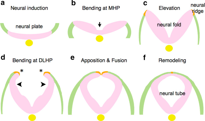
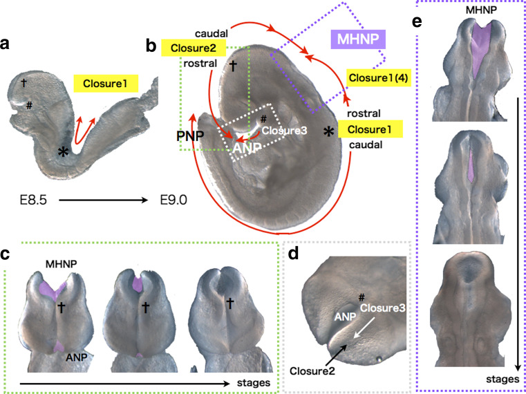
The development of the embryonic brain critically depends on successfully completing cranial neural tube closure (NTC). Failure to properly close the neural tube results in significant and potentially lethal neural tube defects (NTDs). We believe these malformations are caused by disruptions in normal developmental programs such as those involved in neural plate morphogenesis and patterning, tissue fusion, and coordinated cell behaviors. Cranial NTDs include anencephaly and craniorachischisis, both lethal human birth defects. Newly emerging methods for molecular and cellular analysis offer a deeper understanding of not only the developmental NTC program itself but also mechanical and kinetic aspects of closure that may contribute to cranial NTDs. Clarifying the underlying mechanisms involved in NTC and how they relate to the onset of specific NTDs in various experimental models may help us develop novel intervention strategies to prevent NTDs.


Figures

Fig. 1
Fig. 1
Morphological changes of neural plates to neural tube. After neural induction (a), the neural plate bends at MHP (b) and is elevated to form the neural fold (c). Subsequently, flipping of the edges (asterisks) and bending at DLHP occur (d), resulting in apposition and fusion of the edges (e). Remodeling takes place to separate neuroectoderm and surface ectoderm (f). Neuroectoderm (neuroepithelium): pink. Non-neural ectoderm (surface ectoderm): green. Boundary region within non-neural ectoderm: Orange. Notochord: yellow
Fig. 2
Fig. 2
Multiple closures in cranial NTC of mouse embryos. a Bi-directional closure 1 occurs from the cervical region (asterisks) before embryonic turning in an E8.5 ICR embryo. b Schematic representation of multiple closures in an E8.75 ICR embryo. MHNP is closed by caudal closure 2 and rostral closure 1(closure 4), and ANP by caudal closure 2 and closure 3. asterisks: closure 1 start site. Crosses: bi-directional closure 2 start site. Sharp: closure 3 start site. Directions of the closures are shown by red arrows. c Frontal views of closure 2 at MHNP and ANP. d Ventral view of closure 3 at ANP. e Dorsal views of closure 1(4) and 2 at MHNP. Unclosed regions are colored purple
Fig. 3
Fig. 3
Mechanisms of bending at MHP in the cranial regions identified in chicken embryos. a Signals involved in MHP formation, and mechanisms of their actions in chicken cranial region. b PCP signaling links convergent extension with neural plate bending via oriented apical constriction in chicken cranial region. Oriented apical constriction along mediolateral (ML) axis within neuroepithelial cells (actin fibers are shown with red) couples elongation of the neural plate along anteroposterior (AP) axis with its bending along ML axis
Fig. 4
Fig. 4
Schematic illustration of events occurring in the dorsal neural folds during cranial NTC. Cellular behaviors and molecular mechanisms are shown with blue and black fonts, respectively. Neuroectoderm (neuroepithelium): pink. Non-neural ectoderm (surface ectoderm): green. Boundary region within non-neural ectoderm: orange. Boundary cells mediating fusion at tips: red. Cranial neural crest cells (CNC): purple. Head mesenchyme: light blue. Apoptotic dying cells: gray. A cell undergoing division is shown with yellow in surface ectoderm (left)
Fig. 5
Fig. 5
Developmental time-window model for cranial NTC. NTC must be completed by a hypothetical developmental deadline (about somite stage 20), when forces incompatible with NTC may arise. Any perturbation on NTC program could delay NTC. Even when closure is delayed, the embryo can develop without NTDs, as long as NTC can be completed before the deadline (shown as “rescue form delay”). However, if closure is not completed by the deadline, cranial NTC ends in failure to close at the MHNP, resulting in cranial NTDs such as exencephaly

References

    1. Mitchell L. Epidemiology of neural tube defects. Am J Med Genet C Semin Med Genet. 2005;135C:88–94. doi: 10.1002/ajmg.c.30057. - DOI - PubMed
    1. Copp A, Greene N, Murdoch J. The genetic basis of mammalian neurulation. Nat Rev Genet. 2003;4:784–793. doi: 10.1038/nrg1181. - DOI - PubMed
    1. Copp A, Greene N. Genetics and development of neural tube defects. J Pathol. 2010;220:217–230. - PMC - PubMed
    1. Juriloff D, Harris M. Mouse models for neural tube closure defects. Hum Mol Genet. 2000;9:993–1000. doi: 10.1093/hmg/9.6.993. - DOI - PubMed
    1. Beaudin A, Stover P. Folate-mediated one-carbon metabolism and neural tube defects: balancing genome synthesis and gene expression. Birth Defects Res C Embryo Today. 2007;81:183–203. doi: 10.1002/bdrc.20100. - DOI - PubMed

Publication types

LinkOut - more resources