

Di-n-butyl phthalate disrupts the expression of genes involved in cell cycle and apoptotic pathways in mouse ovarian antral follicles
- PMID: 23242528
- PMCID: PMC4434941
- DOI: 10.1095/biolreprod.112.105122
Di-n-butyl phthalate disrupts the expression of genes involved in cell cycle and apoptotic pathways in mouse ovarian antral follicles
Abstract
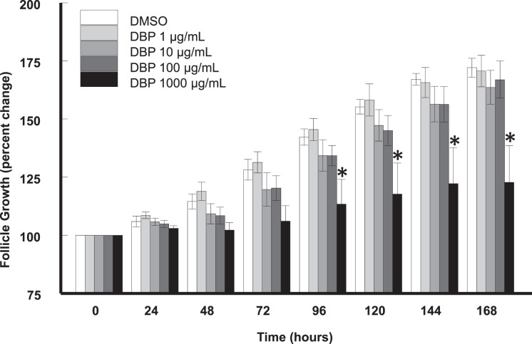
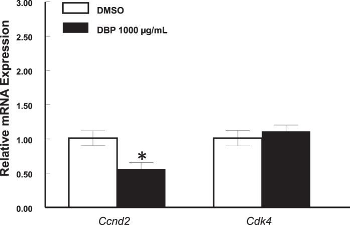
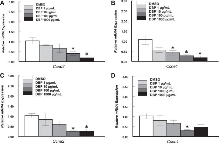
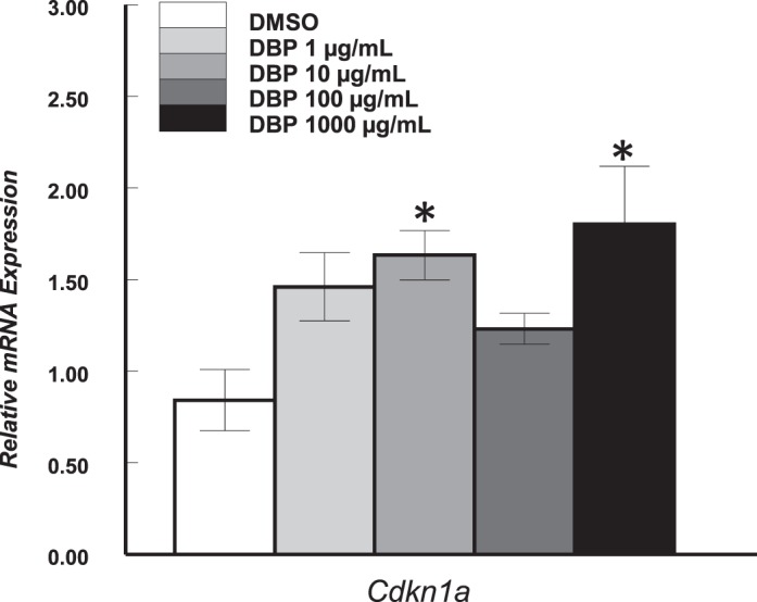
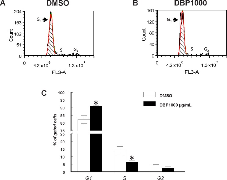
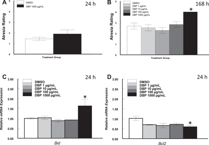
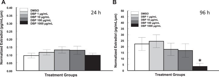
Di-n-butyl phthalate (DBP) is present in many consumer products, such as infant, beauty, and medical products. Several studies have shown that DBP causes reproductive toxicity in rodents, but no studies have evaluated its effects on ovarian follicles. Therefore, we used a follicle culture system to evaluate the effects of DBP on antral follicle growth, cell cycle and apoptosis gene expression, cell cycle staging, atresia, and 17β-estradiol (E(2)) production. Antral follicles were isolated from adult CD-1 mice and exposed to DBP at 1, 10, 100, and 1000 μg/ml for 24 or 168 h. Follicles treated with vehicle or DBP at 1-100 μg/ml grew over time, but DBP at 1000 μg/ml significantly suppressed follicle growth. Regardless of effect on follicle growth, DBP-treated follicles had decreased mRNA for cyclins D2, E1, A2, and B1 and increased p21. Levels of the proapoptotic genes Bax, Bad, and Bok were not altered by DBP treatment, but DBP 1000 μg/ml increased levels of Bid and decreased levels of the antiapoptotic gene Bcl2. DBP-treated follicles contained significantly more cells in G(1) phase, significantly less cells in S, and exhibited a trend for fewer cells in G(2). Although DBP did not affect E(2) production and atresia at 24 h, follicles treated with DBP had reduced levels of E(2) at 96 h and underwent atresia at 168 h. These data suggest that DBP targets antral follicles and alters the expression of cell cycle and apoptosis factors, causes cell cycle arrest, decreases E(2), and triggers atresia, depending on dose.
Figures
Similar articles
-
Effects of in vitro exposure to dibutyl phthalate, mono-butyl phthalate, and acetyl tributyl citrate on ovarian antral follicle growth and viability.Biol Reprod. 2017 May 1;96(5):1105-1117. doi: 10.1095/biolreprod.116.144691. Biol Reprod. 2017. PMID: 28486587 Free PMC article.
-
Di(2-ethylhexyl) phthalate inhibits antral follicle growth, induces atresia, and inhibits steroid hormone production in cultured mouse antral follicles.Toxicol Appl Pharmacol. 2015 Apr 1;284(1):42-53. doi: 10.1016/j.taap.2015.02.010. Epub 2015 Feb 18. Toxicol Appl Pharmacol. 2015. PMID: 25701202 Free PMC article.
-
Effects of an Environmentally Relevant Phthalate Mixture on Cultured Mouse Antral Follicles.Toxicol Sci. 2017 Mar 1;156(1):217-229. doi: 10.1093/toxsci/kfw245. Toxicol Sci. 2017. PMID: 28013214 Free PMC article.
-
Short term exposure to di-n-butyl phthalate (DBP) disrupts ovarian function in young CD-1 mice.Reprod Toxicol. 2015 Jun;53:15-22. doi: 10.1016/j.reprotox.2015.02.012. Epub 2015 Mar 9. Reprod Toxicol. 2015. PMID: 25765776 Free PMC article.
-
Quantitative label-free proteomic analysis of mouse ovarian antral follicles following oral exposure to a human-relevant mixture of three phthalates.Toxicol Sci. 2024 Oct 1;201(2):226-239. doi: 10.1093/toxsci/kfae089. Toxicol Sci. 2024. PMID: 38995844 Free PMC article.
Cited by
-
Single-cell RNA-seq reveals that granulosa cells are a target of phthalate toxicity in the ovary.Toxicol Sci. 2025 Apr 1;204(2):169-180. doi: 10.1093/toxsci/kfaf001. Toxicol Sci. 2025. PMID: 39752319
-
The effects of endocrine-disrupting chemicals on ovarian- and ovulation-related fertility outcomes.Mol Reprod Dev. 2022 Dec;89(12):608-631. doi: 10.1002/mrd.23652. Epub 2022 Nov 19. Mol Reprod Dev. 2022. PMID: 36580349 Free PMC article. Review.
-
Effects of Endocrine-Disrupting Chemicals on the Ovary.Biol Reprod. 2015 Jul;93(1):20. doi: 10.1095/biolreprod.115.130336. Epub 2015 Jun 10. Biol Reprod. 2015. PMID: 26063868 Free PMC article. Review.
-
Perfluorooctanoic Acid Disrupts Ovarian Steroidogenesis and Folliculogenesis in Adult Mice.Toxicol Sci. 2022 Mar 28;186(2):260-268. doi: 10.1093/toxsci/kfac005. Toxicol Sci. 2022. PMID: 35104888 Free PMC article.
-
Developmental programming: mechanisms of early exposure to real-life chemicals in biosolids on offspring ovarian dynamics†.Biol Reprod. 2025 Jun 15;112(6):1229-1242. doi: 10.1093/biolre/ioaf053. Biol Reprod. 2025. PMID: 40121545
References
-
- Center for the Evaluation of Risks to Human Reproduction. NTP-CERHR Expert Panel Report on Di(2-Ethylhexyl) Phthalate Alexandria, VA: Science International, Inc.; 2000.
-
- Third National Report on Human Exposure to Environmental Chemicals NCEH Publication No. 05–0570 Washington, DC: Department of Health and Human Services, Centers for Disease Control and Prevention; 2005.
-
- National Toxicology Program Report on Carcinogens, 8th ed Research Triangle Park, NC: National Toxicology Program; 1998.
-
- Aldyreva MV, Klimova TS, Iziumova AS, Timofeevskaia LA. The effect of phthalate plasticizers on the generative function. Gig Tr Prof Zabol. 1975;19:25–29. - PubMed
Publication types
MeSH terms
Substances
Grants and funding
LinkOut - more resources
Full Text Sources
Other Literature Sources
Research Materials
