. 2013 Mar;12(3):792-6.
doi: 10.1074/mcp.M112.023663. Epub 2012 Dec 13.

Matrix assisted ionization vacuum (MAIV), a new ionization method for biological materials analysis using mass spectrometry

Affiliations

Matrix assisted ionization vacuum (MAIV), a new ionization method for biological materials analysis using mass spectrometry

Ellen D Inutan et al. Mol Cell Proteomics. 2013 Mar.

Abstract

The introduction of electrospray ionization (ESI) and matrix-assisted laser desorption/ionization (MALDI) for the mass spectrometric analysis of peptides and proteins had a dramatic impact on biological science. We now report that a wide variety of compounds, including peptides, proteins, and protein complexes, are transported directly from a solid-state small molecule matrix to gas-phase ions when placed into the vacuum of a mass spectrometer without the use of high voltage, a laser, or added heat. This ionization process produces ions having charge states similar to ESI, making the method applicable for high performance mass spectrometers designed for atmospheric pressure ionization. We demonstrate highly sensitive ionization using intermediate pressure MALDI and modified ESI sources. This matrix and vacuum assisted soft ionization method is suitable for the direct surface analysis of biological materials, including tissue, via mass spectrometry.


Figures

Fig. 1.
Fig. 1.
MAIV mass spectra. A, lysozyme, 14.3 kDa. B, noncovalent complex of lysozyme/penta-N-acetylchitopentaose (PNAP) extracted from the IMS-MS two-dimensional dataset. C, total ion current of A. D, bovine serum albumin, ∼66 kDa. Red numbers indicate the charge states, and blue numbers in the upper right corner of each spectrum provide relative ion abundances.
Fig. 2.
Fig. 2.
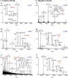
MAIV mass spectra. (I) Positive ion mode: (A) 1 pmol leucine enkephalin (molecular weight (MW) 555 Da), (B) 1 pmol MBP peptide (MW 1833 Da), and (C) 50 fmol ubiquitin (MW 8559 Da). (II) Negative ion mode: (A) ubiquitin, (B) phosphorylated peptide cholecystokinin (10–20) (MW 1332 Da), and (C) hirudin (55–65) peptide (MW 1411 Da).
Fig. 3.
Fig. 3.
MAIV-IMS-MS of a mouse brain tissue section using a commercial vacuum MALDI source with the laser off. A, photograph of application of matrix solution to a section of mouse brain on a glass microscope slide. B, two-dimensional IMS-MS plot of drift time versus m/z of ions showing separation of compound classes by charge, size, and shape of lipids, peptides, and proteins. Insets show isotopic distributions of lipid and protein ions. C, microscopy photograph of the mouse brain tissue before and after ionization. Circled area shows the area on the tissue analyzed in B, and small arrows are included to guide the eye for a better relative comparison of the photographs.
Fig. 4.
Fig. 4.
MAIV-IMS-MS of a solvent-extracted blood spot from a Band-Aid using a commercial atmospheric pressure ESI source with a modified skimmer cone to provide a larger inlet aperture. A, photograph of the device showing the isolation valve in the open position with a glass microscope slide held by the pressure differential against the skimmer opening, with the matrix–blood spot sample exposed to the vacuum initiating ionization; more details on source modifications are provided in supplemental Fig. S1. B, two-dimensional IMS-MS plot of drift time versus m/z of ions showing separation of compound classes by charge, size, and shape: α-globin (MW 15,126 Da) and β-globin (MW 15,866 Da) chains of hemoglobin. Both α and β chains of hemoglobin are detected with MAIV in good ion abundance (mass spectral details in supplemental Fig. S2), something that has been reported (24) to be problematic with ESI (1). Extraction of the mass spectral information from the two-dimensional display in B provides the individual mass spectra of the proteins. C, extracted full range mass spectrum of the β-globin chain.
Fig. 5.
Fig. 5.
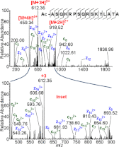
MAIV using the LTQ Velos ESI source to perform ETD fragmentation of the MBP peptide +4 charge state ion (∼m/z 459). Top, total fragment spectrum; bottom, inset m/z 400–900. Sequence coverage is increased relative to previous ETD studies of this peptide (14). Red numbers indicate the charge state, and blue numbers the ion abundance. Sequence coverage is provided (top).

Similar articles

Cited by

References

    1. Fenn J. B., Mann M., Meng C. K., Wong S. F., Whitehouse C. M. (1989) Electrospray ionization for mass spectrometry of large biomolecules. Science 246, 64–71 - PubMed
    1. Tanaka K., Waki H., Ido Y., Akita S., Yoshida Y. (1988) Protein and polymer analyses up to m/z 100 000 by laser ionization time-of-flight mass spectrometry. Rapid Commun. Mass Spectrom. 2, 151–153
    1. Vestal M. L. (2009) Modern MALDI time-of-flight mass spectrometry. J. Mass Spectrom. 44, 303–317 - PubMed
    1. Rosinke B., Strupat K., Hillenkamp F., Rosenbusch J., Dencher N., Kruger U., Galla H. J. (1995) Matrix-assisted laser desorption/ionization mass-spectrometry (MALDI-MS) of membrane-proteins and noncovalent complexes. J. Mass Spectrom. 30, 1462–1468
    1. Bich C., Zenobi R. (2009) Mass spectrometry of large complexes. Curr. Opin. Struct. Biol. 19, 632–639 - PubMed

Publication types